
Integrating Microbial Exopolysaccharides (EPS) with Growth Factors for Enhanced Antimicrobial and Angiogenic Wound Healing
-
Tahmid Anas
Department of Biochemistry and Microbiology, North South University, Basundhara R/A, Dhaka, Bangladesh
Md. Asaduzzaman ShishirDepartment of Microbiology, Dhaka International University, Satarkul, Dhaka, Bangladesh
Kazi Fahmida RahmanDepartment of Biochemistry and Microbiology, North South University, Basundhara R/A, Dhaka, Bangladesh
Kazi Samia PialDepartment of Biochemistry and Microbiology, North South University, Basundhara R/A, Dhaka, Bangladesh
Md. Murshed Hasan SarkarDepartment of Biochemistry and Microbiology, North South University, Basundhara R/A, Dhaka, Bangladesh
Kazi Md. Mostafizur RahmanDepartment of Biochemistry and Microbiology, North South University, Basundhara R/A, Dhaka, Bangladesh
S.M. Bakhtiar ul IslamDepartment of Biochemistry and Microbiology, North South University, Basundhara R/A, Dhaka, Bangladesh
Nayeema BulbulDepartment of Biochemistry and Microbiology, North South University, Basundhara R/A, Dhaka, Bangladesh
Jinath Sultana JimeDepartment of Biochemistry and Microbiology, North South University, Basundhara R/A, Dhaka, Bangladesh
Ashrafus SafaDepartment of Life Sciences, Independent University, Bangladesh, Basundhara R/A, Dhaka, Bangladesh
Md. Fakruddin
Department of Biochemistry and Microbiology, North South University, Basundhara R/A, Dhaka, Bangladesh
| Received 15 Jan, 2026 |
Accepted 22 Jul, 2026 |
Published 30 Sep, 2026 |
Chronic wounds, including diabetic foot ulcers, venous leg ulcers, and pressure ulcers, represent a major clinical challenge due to persistent microbial infection, prolonged inflammation, impaired angiogenesis, and delayed tissue regeneration. Conventional wound care strategies often fail to address these interconnected pathological factors simultaneously, resulting in poor healing outcomes and high recurrence rates. Microbial exopolysaccharides (EPS) have emerged as promising biomaterials for wound healing due to their biocompatibility, moisture-retention capacity, antimicrobial barrier properties, and extracellular matrix-mimicking structure. In parallel, growth factors such as vascular endothelial growth factor, platelet-derived growth factor, fibroblast growth factor, and epidermal growth factor play essential roles in regulating angiogenesis, cell proliferation, and tissue remodeling. However, direct application of growth factors is limited by rapid degradation, poor localization, and uncontrolled release in protease-rich chronic wound environments. Integrating growth factors within EPS-based matrices offers a multifunctional therapeutic strategy that combines antimicrobial protection, structural support, and sustained pro-regenerative signaling. The EPS-GF composite systems enable localized and controlled growth factor delivery, promote neovascularization, enhance extracellular matrix deposition, and reduce microbial burden at the wound site. Preclinical studies consistently demonstrate accelerated wound closure and improved tissue quality using these integrated platforms compared to conventional dressings or single-component therapies. This review primarily focuses on bacteria-derived EPS, with limited discussion of selected fungal EPS relevant to wound healing applications., Also discusses their biological roles, strategies for their integration, mechanistic insights into their synergistic action, and recent preclinical advances. Despite their strong therapeutic potential, challenges related to large-scale production, growth factor stability, regulatory approval, clinical translation remain and must be addressed to enable widespread clinical adoption. Overall, EPS-GF composites represent a next-generation approach for advanced wound management with strong potential for clinical translation.
| Copyright © 2026 Anas et al. This is an open-access article distributed under the Creative Commons Attribution License, which permits unrestricted use, distribution, and reproduction in any medium, provided the original work is properly cited. |
INTRODUCTION
Chronic wounds, including diabetic foot ulcers, pressure injuries, and severe burn lesions, remain a major clinical problem due to delayed healing, high infection risk, and substantial healthcare costs1-7. Unlike acute wounds that progress through orderly healing phases, chronic wounds frequently remain in a prolonged inflammatory state marked by excessive protease activity, elevated reactive oxygen species, and impaired angiogenesis1,2. Chronic wounds are characterized by persistent microbial colonization, excessive inflammatory responses, and disrupted angiogenic signaling, which collectively delay tissue regeneration and wound closure8. Microbial colonization and host microbiota dysregulation can significantly influence inflammation and tissue repair outcomes in chronic wound environments9. Persistent microbial colonization, particularly by biofilm-forming pathogens, further sustains inflammation and restricts cellular migration, creating a vicious cycle that conventional therapies rarely resolve3. Standard wound dressings provide moisture and protection but do not address these underlying biological dysfunctions4.
Microbial exopolysaccharides (EPS) have emerged as promising biomaterials for chronic wound management due to their biocompatibility, natural origin, and multifunctionality5,6. The EPS forms hydrated hydrogel networks that mimic the extracellular matrix (ECM), supporting cell adhesion, migration, and proliferation while maintaining a moist wound environment. Several EPS, including bacterial cellulose, xanthan, hyaluronic acid, and dextran derivatives, also exhibit intrinsic antimicrobial activity through inhibition of bacterial adhesion, biofilm disruption, or immune modulation5,6, making them effective structural and protective scaffolds.
Growth factors (GF), such as VEGF, PDGF, FGF, and EGF, are central regulators of angiogenesis, fibroblast activity, extracellular matrix deposition, and epithelialization7. However, their clinical use is limited by rapid degradation, short half-life, and uncontrolled release. Incorporation of growth factors into EPS-based hydrogels enables sustained and localized delivery while protecting bioactivity and maintaining an ECM-like microenvironment conducive to neovascularization and tissue repair6,7.
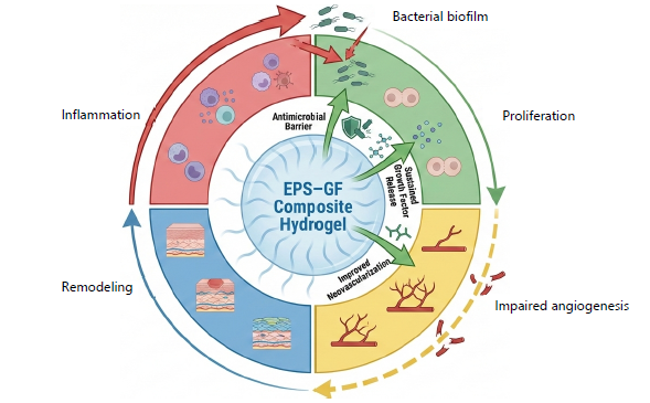
Chronic wounds persist through interconnected processes of infection, inflammation, and poor vascularization. As illustrated in Fig. 1, chronic wounds are characterized by a self-perpetuating vicious cycle involving persistent bacterial biofilm formation, prolonged inflammation, impaired angiogenesis, and delayed tissue remodeling. The schematic highlights how EPS-GF composite hydrogels intervene in this cycle by providing an antimicrobial barrier, enabling sustained growth factor release, promoting neovascularization, and supporting cellular proliferation and tissue regeneration. The EPS-GF composites are designed to disrupt this cycle by integrating antimicrobial protection, controlled pro-regenerative signaling, and structural support. This review examines the biological basis of wound healing, the functional roles of microbial EPS and growth factors, recent advances in EPS-GF composite systems, and strategies for their integration into multifunctional wound therapies to support clinical translation.
To ensure a structured and comprehensive narrative review, a literature search was conducted focusing on microbial exopolysaccharide (EPS)-based growth factor delivery systems for wound healing applications. Relevant studies were identified using electronic databases, including PubMed, Scopus, and Web of Science. Additional articles were retrieved through manual screening of reference lists from selected publications to ensure broader coverage of relevant literature.
Search terms were formulated using combinations of keywords and Boolean operators, including “microbial exopolysaccharides”, “bacterial cellulose”, “dextran”, “xanthan”, “hyaluronic acid”, “growth factor delivery”, “VEGF”, “PDGF”, “FGF”, “EGF”, “chronic wounds”, and “angiogenesis”. These terms were used to capture studies addressing both the antimicrobial and regenerative roles of EPS-based systems.
|
Peer-reviewed articles published in English between 2000 and 2025 were included. Studies were selected based on their relevance to microbial-derived EPS materials in wound healing, particularly those involving growth factor incorporation and reporting biological or therapeutic outcomes. Duplicate records, conference abstracts, non-peer-reviewed sources, and studies unrelated to wound healing were excluded.
Both in vitro and in vivo preclinical studies were considered, along with relevant clinical reports where applicable. Study selection was performed through title and abstract screening, followed by full-text evaluation based on relevance to the review scope. Extracted information was organized according to EPS type, growth factor involved, delivery strategy, biological performance, and translational relevance. The findings were synthesized qualitatively to highlight key mechanistic insights and recent advancements in the field10.
MICROBIAL EXOPOLYSACCHARIDES (EPS) IN WOUND HEALING
Microbial exopolysaccharides (EPS) are high-molecular-weight polysaccharides produced by bacteria and fungi that have gained increasing attention in wound healing due to their multifunctional properties11,12. The EPS readily forms hydrated hydrogel networks that mimic the extracellular matrix (ECM), providing structural support, maintaining a moist wound environment, and facilitating cellular infiltration necessary for tissue repair13. Microbial exopolysaccharides provide a hydrated three-dimensional matrix that mimics native extracellular matrix architecture while simultaneously acting as a protective antimicrobial barrier8. Their physicochemical properties, including viscosity, mechanical strength, and degradation rate, can be tuned by modifying monosaccharide composition and molecular architecture14.
There are many kinds of EPS investigated for wound healing, including hyaluronic acid (HA), dextran, xanthan, bacterial cellulose (BC), alginate, and gellan gum. HA regulates inflammation, promotes angiogenesis, and supports fibroblast proliferation while maintaining hydration15. Dextran-based hydrogels exhibit good biocompatibility with mild antimicrobial effects that reduce infection risk16. Xanthan functions as a scaffold and viscosity modifier with inhibitory effects on bacterial growth17. The BC is distinguished by its high tensile strength, nanofibrillar structure, and excellent water retention, making it an effective regenerative scaffold16. Alginate and gellan gum form ionically crosslinked hydrogels that maintain moisture and support cell adhesion and controlled delivery of bioactive agents17.
| Table 1: | Selected microbial eps and their dual functions in wound healing | |||
| EPS type | Source | Primary antimicrobial mechanism | Pro-regenerative function | Suitability for GF incorporation | Key references |
| Hyaluronic acid | Streptococcus spp. | Inhibits bacterial adhesion; hydration barrier | Promotes angiogenesis and cell migration | Excellent | Wan et al.13 and Litwiniuk et al.15 |
| Bacterial cellulose | Komagataeibacter spp. | Physical microbial barrier | Supports angiogenesis and granulation | Excellent | Arslan et al.6 and Boateng et al.19 |
| Dextran | Leuconostoc spp. | Reduces biofilm formation | Enhances fibroblast activity | Good | Portela et al.16 |
| Xanthan gum | Xanthomonas campestris | Limits microbial colonization | Maintains moist wound environment | Moderate | Bharathi and Lee11 and Lee and Mooney17 |
| Alginate (microbial) | Azotobacter spp. | Gel barrier against microbes | Supports cell proliferation | Good | Boateng et al.19 and Tan et al.20 |
| Pullulan | Aureobasidium pullulans | Prevents bacterial attachment | Enhances epithelialization | Moderate | Boateng et al.19 |
The EPS show antimicrobial activity by limiting microbial adhesion, disrupting biofilm formation, and modulating host immune responses11,13. Additionally, microbial-derived biotherapeutics such as next-generation probiotics may indirectly support wound healing by modulating host immunity and inflammation18. The BC hydrogels, for example, restrict colonization by Staphylococcus aureus and Pseudomonas aeruginosa, while xanthan and dextran derivatives indirectly suppress bacterial growth by enhancing host defenses16,17,19.
Beyond infection control, EPS actively supports angiogenesis and tissue regeneration. HA enhances endothelial cell migration and neovascularization15, while BC and gellan gum provide scaffolding for fibroblast attachment, collagen deposition, and granulation tissue formation19,20.
The EPS hydrogels are also well suited for growth factor loading. Hydrophilic networks can entrap VEGF, PDGF, FGF, and EGF, enabling controlled and sustained release13. Release behavior can be regulated through crosslinking density and degradation rate14, while covalent conjugation in BC or modified HA further stabilizes growth factors and prolongs bioactivity15.
As summarized in Table 1, microbial EPS combine antimicrobial activity, pro-regenerative function, and compatibility with growth factor incorporation. Chemical or enzymatic modifications such as sulfation, acetylation, or crosslinking further enhance mechanical properties and bioactivity16. The EPS can also act as carriers for additional bioactive agents, including antimicrobial peptides or nanoparticles, enabling multifunctional wound dressings16.
From my perspective, microbial EPS represent a versatile and adaptable platform for chronic wound healing, offering structural support, antimicrobial protection, and effective growth factor delivery beyond what conventional dressings can provide.
GROWTH FACTORS IN WOUND HEALING
Growth factors (GFs) are important signaling molecules that control significant steps in tissue repair, such as cell proliferation, migration, angiogenesis, and extracellular matrix deposition21,22. They are main controllers of both acute and chronic wound healing, coordinating cellular responses and tissue regeneration. Some of the most important growth factors involved in wound repair include Vascular Endothelial Growth Factor (VEGF), Platelet-Derived Growth Factor (PDGF), Fibroblast Growth Factor (FGF), and Epidermal Growth Factor (EGF), which act through complementary mechanisms23. However the protease-rich microenvironment of chronic wounds significantly reduces growth factor bioavailability, limiting their therapeutic efficacy when administered directly8.
The VEGF drives angiogenesis by stimulating endothelial cell proliferation and capillary formation, restoring vascular supply in ischemic wounds24. It is a critical biological process for tissue regeneration in chronic wound environments25. The PDGF promotes fibroblast recruitment, proliferation, and matrix production, supporting granulation tissue formation26. The FGF exhibits broad mitogenic effects on fibroblasts, keratinocytes, and endothelial cells, facilitating both neovascularization and epithelialization27, while EGF accelerates wound closure by enhancing keratinocyte migration and proliferation.
Despite their therapeutic potential, free growth factors show limited clinical efficacy due to rapid enzymatic degradation, short half-life, and instability within the chronic wound microenvironment24,26. Uncontrolled or burst release can also cause off-target effects such as fibrosis or abnormal angiogenesis, often requiring repeated or high-dose administration27.
To overcome these limitations, controlled delivery strategies have been developed, including physical entrapment in hydrogels, covalent conjugation to polymeric scaffolds, and affinity-based binding via heparin or related interactions21,22. These approaches improve growth factor stability and enable sustained, localized release while preserving bioactivity23.
Chronic wounds are also marked by higher protease activity and persistent inflammation, which reduce growth factor bioavailability24. Oxidative stress and excessive reactive oxygen species further disrupt angiogenic signaling and delay healing processes28. Incorporation into EPS-based hydrogels provides a protective, ECM-like reservoir that preserves growth factor activity and allows modulation of release kinetics through network density, porosity, and degradation rate26. Sequential or staged release can better mimic physiological healing, with early PDGF-driven fibroblast recruitment followed by VEGF-mediated angiogenesis and EGF-induced epithelialization27.
Consistent preclinical evidence shows that growth factor-loaded hydrogels outperform free growth factor delivery, significantly enhancing angiogenesis, granulation tissue formation, fibroblast proliferation, and collagen deposition in diabetic wound models21,27.
In conclusion, while growth factors are essential for wound healing, their clinical application is limited by instability and rapid degradation. The EPS-based hydrogels enable controlled, localized delivery and sustained bioactivity, forming the foundation for effective EPS-GF composite systems in chronic wound therapy.
EPS-GF COMPOSITE STRATEGIES
The integration of growth factors (GFs) into microbial exopolysaccharide (EPS) matrices represents a sophisticated approach to addressing the dual challenges of infection control and tissue regeneration in chronic wounds29. Incorporating growth factors within biopolymeric matrices enhances their localized retention, promotes sustained release kinetics, and improves angiogenic and regenerative outcomes8. The EPS-GF composites leverage the structural and antimicrobial properties of EPS while providing controlled, localized delivery of bioactive growth factors30. Several strategies have been developed to incorporate growth factors into EPS hydrogels, primarily categorized into physical entrapment, covalent conjugation, and affinity-based binding. Each method offers distinct advantages and limitations.
Physical entrapment: Physical entrapment involves incorporating growth factors directly within the hydrogel matrix during scaffold formation. The EPS network retains the growth factors and allows gradual diffusion over time, maintaining bioactivity without chemical modification31. This method is straightforward and preserves protein structure but may lead to an initial burst release, potentially limiting sustained therapeutic efficacy. The EPS types such as bacterial cellulose, hyaluronic acid, and alginate are well-suited for physical entrapment due to their high water content, mechanical stability, and biocompatibility.
| Table 2: | Comparison of growth factor integration strategies in EPS matrices | |||
| Method | GF examples | Release kinetics | Advantages | Limitations | Key references |
| Physical entrapment | VEGF, FGF | Burst+diffusion-controlled | Simple, preserves bioactivity | Poor long-term control | Li et al.22 and Peppas et al.31 |
| Covalent conjugation | VEGF, PDGF | Sustained, minimal burst | High stability, localized delivery | Possible loss of bioactivity | Richardson et al.32 |
| Affinity-based binding | VEGF, FGF | Reversible, controlled | Mimics ECM regulation | Complex fabrication | Ruel-Gariépy and Leroux33 |
| Microparticle- in-hydrogel | VEGF, EGF | Multi-phase release | Excellent GF protection | Fabrication complexity | Zhang et al.29 and Tonnesen et al.34 |
| VEGF: Vascular Endothelial Growth Factor (primarily responsible for stimulating the growth of new blood vessels), FGF: Fibroblast Growth Factor (involved in angiogenesis, wound healing, and embryonic development). PDGF: Platelet-Derived Growth Factor (plays a significant role in blood vessel formation and cell growth) and EGF: Epidermal Growth Factor (plays an important role in the regulation of cell growth, proliferation, and differentiation) | |||||
Covalent conjugation: Covalent conjugation chemically links growth factors to functional groups on EPS polymer chains. This strategy enhances growth factor stability, reduces enzymatic degradation, and provides a predictable, sustained release profile32. Functional groups such as amines or carboxyls on EPS can be modified to bind VEGF or PDGF, stabilizing the protein within the hydrogel. While covalent conjugation offers superior control over release kinetics, it requires careful optimization to avoid altering growth factor bioactivity. Chemically modified EPS, including thiolated hyaluronic acid or carboxymethyl dextran, have been successfully employed for covalent immobilization of growth factors32.
Affinity-based binding: Affinity-based binding utilizes specific interactions between growth factors and binding motifs incorporated into the EPS matrix. Heparin-like domains or engineered binding sites can reversibly capture growth factors such as VEGF and FGF, allowing controlled release in response to environmental signals33. This approach mimics natural extracellular matrix regulation, enabling temporal and spatial presentation of growth factors without covalent modification. It is particularly useful for sequential or gradient-based delivery, aligning with the wound healing process.
Comparison of strategies: Table 2 summarizes these EPS-GF integration strategies, including method, representative growth factors, release kinetics, advantages, and limitations. Physical entrapment is simple and preserves bioactivity but can result in burst release. Covalent conjugation ensures long-term stability but requires chemical modification. Affinity-based binding allows reversible, temporally controlled release but is more complex to engineer. Selection of the appropriate strategy depends on the wound type, desired release profile, and specific EPS properties.
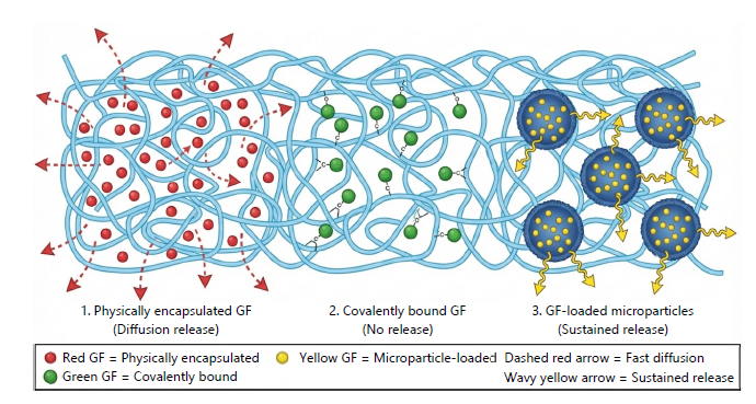
Design considerations: Hydrogel performance depends on EPS type, crosslinking density, porosity, degradation rate, and mechanical properties29. Hydrogels must balance structural integrity with flexibility to conform to irregular wound surfaces while maintaining hydration. Combining multiple EPS types or blending with secondary polymers can optimize scaffold properties, enhancing growth factor retention, antimicrobial activity, and tissue regeneration potential30. Figure 2 illustrates representative EPS-GF hydrogel designs: (1) Physically entrapped growth factors, (2) Covalently linked growth factors, and (3) growth factors incorporated via microparticles or affinity domains within the EPS matrix. The figure highlights how integration strategies influence spatial distribution, release kinetics, and cellular interactions at the wound site. Preclinical studies have demonstrated the efficacy of EPS-GF composites in accelerating wound closure and promoting angiogenesis. The VEGF-loaded bacterial cellulose hydrogels enhance neovascularization and granulation tissue formation, while PDGF-incorporated hyaluronic acid scaffolds promote fibroblast proliferation and epithelialization more effectively than free growth factor application29,30. Furthermore, EPS-GF composites can be engineered for stimuli-responsive release. pH-sensitive or enzyme-responsive hydrogels can selectively release growth factors in response to wound-specific cues, enhancing therapeutic precision33. Overall, EPS-GF composites offer a multifunctional platform capable of simultaneously providing antimicrobial protection, sustained growth factor delivery, and structural support for tissue regeneration. Careful selection of EPS type and incorporation strategy allows the design of scaffolds tailored to specific wound types, maximizing therapeutic efficacy while minimizing systemic exposure and complications.
|
MECHANISTIC INSIGHTS AT THE WOUND BED
The EPS-GF composites work by combining antimicrobial protection, structural support, and controlled growth factor delivery at the wound site34. Chronic wounds often suffer from bacterial colonization, biofilm formation, and poor angiogenesis, which hinder healing. The EPS hydrogels act as a physical barrier, preventing bacterial adhesion and biofilm formation, while maintaining a hydrated environment that supports cell migration35. Their nanofibrillar or crosslinked structure also improves scaffold porosity and mechanical strength, aiding fibroblast and endothelial cell infiltration and extracellular matrix deposition.
Sustained release of growth factors ensures continuous signaling to cells. The VEGF promotes endothelial proliferation, migration, and tube formation, supporting neovascularization34. The PDGF and FGF recruit fibroblasts, enhance collagen synthesis, and stimulate epithelial proliferation. By controlling release kinetics, EPS-GF composites maintain effective concentrations over time, addressing the low natural growth factor levels in chronic wounds.
The EPS matrices can be engineered to respond to environmental cues. The pH-sensitive hydrogels release growth factors in acidic wound conditions, while enzyme-responsive scaffolds degrade in the presence of wound-specific proteases, triggering local GF release35. This targeted delivery enhances precision and efficacy.
At the cellular level, EPS-GF scaffolds reduce bacterial load and inflammation, allowing keratinocytes, fibroblasts, and endothelial cells to proliferate efficiently. Embedded growth factors stimulate angiogenesis and extracellular matrix deposition, accelerating granulation tissue formation. This synergistic effect interrupts the chronic wound cycle, promoting re-epithelialization, neovascularization, and functional tissue repair34.
|
| Table 3: | Summary of recent preclinical studies using EPS-GF composites | |||
| EPS type | Growth factor | Wound model | Key outcome | Key references |
| Bacterial cellulose | VEGF | Diabetic rat wound | Enhanced angiogenesis and faster closure | Schultz and Wysocki37 |
| Hyaluronic acid | PDGF | Full-thickness rat wound | Increased fibroblast proliferation | Schultz and Wysocki37 |
| Xanthan gum | FGF | Ischemic wound | Improved granulation tissue | Zhang et al.29 |
| Dextran | VEGF | Infected wound | Reduced bacterial load, better vascularization | Jayakumar et al.38 |
| Chitosan-EPS blend | EGF | Diabetic mouse wound | Accelerated re-epithelialization | Boateng et al.19 |
| Alginate-EPS composite | VEGF | Chronic wound | Sustained GF release, higher capillary density | Ruel-Gariépy and Leroux33 |
| VEGF: Vascular Endothelial Growth Factor (primarily responsible for stimulating the growth of new blood vessels), FGF: Fibroblast Growth Factor (involved in angiogenesis, wound healing, and embryonic development), PDGF: Platelet-Derived Growth Factor (plays a significant role in blood vessel formation and cell growth) and EGF: Epidermal Growth Factor (plays an important role in the regulation of cell growth, proliferation, and differentiation) | ||||
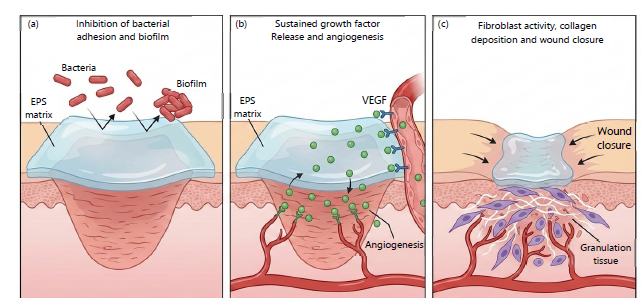
Figure 3 illustrates this mechanism: (A) EPS inhibits bacterial adhesion and biofilms, (B) growth factors like VEGF stimulate angiogenesis, and (C) fibroblast activity drives collagen deposition and wound closure.
In summary, EPS-GF composites provide antimicrobial protection, sustained growth factor signaling, and structural support, overcoming the main limitations of conventional dressings and free growth factor therapies34-36.
PRECLINICAL AND EXPERIMENTAL EVIDENCE OF EPS-GF COMPOSITES
Recent preclinical studies demonstrate the efficacy of EPS-GF composites in chronic wound models, particularly diabetic, ischemic, and infected wounds37. In diabetic rodent models, VEGF-loaded bacterial cellulose hydrogels significantly enhanced angiogenesis, granulation tissue formation, and wound closure compared to free VEGF or EPS alone37. Hyaluronic acid-based hydrogels incorporating PDGF or FGF further improved fibroblast proliferation, collagen deposition, and re-epithelialization. Importantly, the intrinsic antimicrobial properties of EPS reduced bacterial colonization and biofilm formation, creating a favourable environment for tissue regeneration38. Advanced EPS-GF systems have also been engineered to respond to wound-specific cues such as pH or protease activity, enabling targeted and controlled growth factor release. Preclinical investigations demonstrate that biomaterial-based growth factor delivery systems significantly accelerate wound closure and improve tissue remodeling compared to conventional treatments8. Collectively, these findings confirm that EPS-GF composites provide synergistic antimicrobial and pro-regenerative effects, supporting their potential as next-generation wound dressingsin Table 3.
FUTURE PERSPECTIVES AND CHALLENGES
The EPS-GF composites represent a promising strategy for chronic wound management by integrating the antimicrobial and structural properties of microbial exopolysaccharides (EPS) with the regenerative functions of growth factors (GFs)3. Although preclinical studies demonstrate clear therapeutic benefits, several challenges must be addressed to enable clinical translation.
A major limitation is the scalability and standardization of EPS production. Variations in microbial strains, fermentation conditions, and purification processes can affect polymer structure, mechanical properties, and growth factor release behavior3. Improved bioprocess control and reproducible modification strategies are therefore essential for clinical-grade EPS manufacturing.
Growth factor stability, cost, and regulatory concerns also remain critical barriers. While EPS matrices enhance GF protection, large-scale use of recombinant proteins raises issues related to immunogenicity, safety, and batch consistency39. Strategies such as lower-dose combinations of multiple GFs or the use of more stable engineered analogs may help mitigate these challenges.
Stimuli-responsive EPS-GF hydrogels offer a promising direction, enabling growth factor release in response to wound-specific cues such as pH or enzymatic activity40. In addition, multifunctional composites incorporating antimicrobial peptides or nanoparticles may further enhance therapeutic efficacy. Future wound therapies may increasingly rely on microorganism-derived novel bioactive compounds as alternative regenerative and antimicrobial agents41. However, clinical validation remains limited, highlighting the need for systematic human trials evaluating safety, dosing, and long-term outcomes.
Overall, advancing EPS-GF composites toward clinical use will require standardized production, cost-effective design, and rigorous clinical evaluation. Addressing these challenges may enable the development of next-generation wound dressings capable of improving outcomes in complex and chronic wounds.
CONCLUSION
Microbial EPS and growth factors (GFs) act synergistically to support chronic wound healing. By merging EPS’s antimicrobial and structural support with GFs’ angiogenic and regenerative effects, EPS-GF composites tackle the multiple barriers that impair tissue repair. Preclinical studies demonstrate that these composites accelerate wound closure, enhance angiogenesis, promote fibroblast proliferation, and reduce infection risk. The EPS scaffolds can be tailored for physical entrapment, covalent conjugation, or affinity-based binding of growth factors, optimizing release kinetics, preserving protein bioactivity, and supporting cellular infiltration and tissue deposition. This synergy disrupts the chronic wound cycle of inflammation, microbial colonization, and poor vascularization. Future directions include stimuli-responsive hydrogels, multifunctional scaffolds with additional bioactive agents, and scalable, standardized EPS production. Overcoming challenges such as growth factor stability, cost, and clinical translation will be essential. Personalized EPS-GF therapies guided by wound-specific monitoring could further improve healing outcomes. Overall, EPS-GF composites provide antimicrobial protection, sustained growth factor delivery, and extracellular matrix-like structural support, representing a next-generation approach to chronic wound care that can enhance healing, reduce complications, and improve overall patient outcomes.
SIGNIFICANCE STATEMENT
This review highlights microbial exopolysaccharide (EPS)-growth factor (GF) composites as a promising multifunctional strategy for chronic wound healing. By combining the antimicrobial and structural properties of EPS with the regenerative activity of growth factors, these systems enable controlled delivery, enhanced angiogenesis, and improved tissue repair. Overall, EPS-GF platforms offer a next-generation approach to overcome key limitations of conventional wound therapies.
REFERENCES
- Singer, A.J. and R.A.F. Clark, 1999. Cutaneous wound healing. N. Engl. J. Med., 341: 738-746.
- Guo, S. and L.A. DiPietro, 2010. Factors affecting wound healing. J. Dent. Res., 89: 219-229.
- Mustoe, T.A., K.O. Shaughnessy and O. Kloeters, 2006. Chronic wound pathogenesis and current treatment strategies: A unifying hypothesis. Plast. Reconstructive Surg., 117: 35S-41S.
- Falanga, V., 2005. Wound healing and its impairment in the diabetic foot. Lancet, 366: 1736-1743.
- Freitas, F., V.D. Alves and M.A.M. Reis, 2011. Advances in bacterial exopolysaccharides: From production to biotechnological applications. Trends Biotechnol., 29: 388-398.
- Arslan, N.P., T. Orak, A. Ozdemir, R. Altun and N. Esim et al., 2024. Polysaccharides and peptides with wound healing activity from bacteria and fungi. J. Basic Microbiol., 64.
- Barrientos, S., O. Stojadinovic, M.S. Golinko, H. Brem and M. Tomic-Canic, 2008. Growth factors and cytokines in wound healing. Wound Repair Regener., 16: 585-601.
- Fakruddin, M., M.J. Prima, T. Chowdhury, U.T. Ferdous, J. Afroz and M.A. Shishir, 2024. Nature’s tiny chemists: Microorganisms as sources of next-gen active pharmaceutical ingredients (APIs). J. Primeasia, 5.
- Sanam, M., C.F.T.Z. Hossain, Tahmida Binte Hyder, R. Tarannum and J.F. Oishi et al., 2025. Bridging two worlds: Host microbiota crosstalk in health and dysregulation. Innate Immun., 31.
- Page, M.J., J.E. McKenzie, P.M. Bossuyt, I. Boutron and T.C. Hoffmann et al., 2021. The PRISMA 2020 statement: An updated guideline for reporting systematic reviews. BMJ, 372.
- Bharathi, D. and J. Lee, 2025. Polysaccharide-based nanomaterials and hydrogels for antibiofilm and wound healing applications. Int. J. Biol. Macromol., 320.
- Flemming, H.C. and J. Wingender, 2010. The biofilm matrix. Nat. Rev. Microbiol., 8: 623-633.
- Hamidi, M., O.V. Okoro, K. Rashidi, M.S. Salami, R.M. Seveiri, H. Samadian and A. Shavandi, 2022. Evaluation of two fungal exopolysaccharides as potential biomaterials for wound healing applications. World J. Microbiol. Biotechnol., 39.
- Ahmed, E.M., 2015. Hydrogel: Preparation, characterization, and applications: A review. J. Adv. Res., 6: 105-121.
- Litwiniuk, M., A. Krejner and T. Grzela, 2016. Hyaluronic acid in inflammation and tissue regeneration. Wounds, 28: 78-88.
- Portela, R., C.R. Leal, P.L. Almeida and R.G. Sobral, 2019. Bacterial cellulose: A versatile biopolymer for wound dressing applications. Microb. Biotechnol., 12: 586-610.
- Lee, K.Y. and D.J. Mooney, 2001. Hydrogels for tissue engineering. Chem. Rev., 101: 1869-1880.
- Fakruddin, M., M.A. Shishir, Z. Yousuf and M.S.S. Khan, 2022. Next-generation probiotics-the future of biotherapeutics. Microb. Bioactives, 5: 156-163.
- Boateng, J.S., K.H. Matthews, H.N.E. Stevens and G.M. Eccleston, 2008. Wound healing dressings and drug delivery systems: A review. J. Pharm. Sci., 97: 2892-2923.
- Tan, H., C.R. Chu, K.A. Payne and K.G. Marra, 2009. Injectable in situ forming biodegradable chitosan-hyaluronic acid based hydrogels for cartilage tissue engineering. Biomaterials, 30: 2499-2506.
- Park, J.W., S.R. Hwang and I.S. Yoon, 2017. Advanced growth factor delivery systems in wound management and skin regeneration. Molecules, 22.
- Li, Ji., J. Chen and R. Kirsner, 2007. Pathophysiology of acute wound healing. Clin. Dermatol., 25: 9-18.
- Ahmed, T.A.E., E.V. Dare and M. Hincke, 2008. Fibrin: A versatile scaffold for tissue engineering applications. Tissue Eng. Part B: Rev., 14: 199-215.
- Martin, P., 1997. Wound healing--Aiming for perfect skin regeneration. Science, 276: 75-81.
- Fakruddin, M., Z. Chowdhury, S.U.A. Shibly, S.M. Bakhtiar Ul Islam and J.S. Jime et al., 2024. Microbial avengers: How microorganisms drive angiogenesis for good and bad? J. Angiotherapy, 8: 1-12.
- Friedl, P. and K. Wolf, 2003. Tumour-cell invasion and migration: Diversity and escape mechanisms. Nat. Rev. Cancer, 3: 362-374.
- Veith, A.P., K. Henderson, A. Spencer, A.D. Sligar and A.B. Baker, 2019. Therapeutic strategies for enhancing angiogenesis in wound healing. Adv. Drug Delivery Rev., 146: 97-125.
- Fakruddin, M., M.A. Shishir, K.R. Mouree and S.S. Khan, 2022. Environmental and physiological angiogenesis in causing CVD with oxidative pattern. J. Angiotherapy, 6: 663-667.
- Zhang, B., Z. Huang, T. Guo, J. Jing and Y. Dang, 2025. Accelerating infectious wound healing through bacterial cellulose/Ag composite film enriched with GM-CSF. Sci. Rep., 15.
- Ahuja, V., A.K. Bhatt, J.R. Banu, V. Kumar, G. Kumar, Y.H. Yang and S.K. Bhatia, 2023. Microbial exopolysaccharide composites in biomedicine and healthcare: Trends and advances. Polymers, 15.
- Peppas, N.A., J.Z. Hilt, A. Khademhosseini and R. Langer, 2006. Hydrogels in biology and medicine: From molecular principles to bionanotechnology. Adv. Mater., 18: 1345-1360.
- Richardson, T.P., M.C. Peters, A.B. Ennett and D.J. Mooney, 2001. Polymeric system for dual growth factor delivery. Nat. Biotechnol., 19: 1029-1034.
- Ruel-Gariépy, E. and J.C. Leroux, 2004. In situ-forming hydrogels-review of temperature-sensitive systems. Eur. J. Pharm. Biopharm., 58: 409-426.
- Tonnesen, M.G., X. Feng and R.A.F. Clark, 2000. Angiogenesis in wound healing. J. Invest. Dermatol. Symp. Proc., 5: 40-46.
- Costerton, J.W., P.S. Stewart and E.P. Greenberg, 1999. Bacterial biofilms: A common cause of persistent infections. Science, 284: 1318-1322.
- Jones, V., J.E. Grey and K.G. Harding, 2006. Wound dressings. BMJ, 332: 777-780.
- Schultz, G.S. and A. Wysocki, 2009. Interactions between extracellular matrix and growth factors in wound healing. Wound Repair Regener., 17: 153-162.
- Jayakumar, R., M. Prabaharan, P.T.S. Kumar, S.V. Nair and H. Tamura, 2011. Biomaterials based on chitin and chitosan in wound dressing applications. Biotechnol. Adv., 29: 322-337.
- Gurtner, C.G., S. Werner, Y. Barrandon and M.T. Longaker, 2008. Wound repair and regeneration. Nature, 453: 314-321.
- Psarrou, M., A. Mitraki, M. Vamvakaki and C. Kokotidou, 2023. Stimuli-responsive polysaccharide hydrogels and their composites for wound healing applications. Polymers, 15.
- Amin, T., A.B. Karim, I.I. Oyshe, A. Hossain and T. Karim et al., 2023. Unlocking nature’s treasure trove: Exploring microorganisms for novel bioactives. J. Angiotherapy, 7.
How to Cite this paper?
APA-7 Style
Anas,
T., Shishir,
M.A., Rahman,
K.F., Pial,
K.S., Hasan Sarkar,
M.M., Mostafizur Rahman,
K.M., Bakhtiar ul Islam,
S.M., Bulbul,
N., Jime,
J.S., Safa,
A., Fakruddin,
M. (2026). Integrating Microbial Exopolysaccharides (EPS) with Growth Factors for Enhanced Antimicrobial and Angiogenic Wound Healing. Trends in Pharmacology and Toxicology, 2(2), 130-140. https://doi.org/10.21124/tpt.2026.130.140
ACS Style
Anas,
T.; Shishir,
M.A.; Rahman,
K.F.; Pial,
K.S.; Hasan Sarkar,
M.M.; Mostafizur Rahman,
K.M.; Bakhtiar ul Islam,
S.M.; Bulbul,
N.; Jime,
J.S.; Safa,
A.; Fakruddin,
M. Integrating Microbial Exopolysaccharides (EPS) with Growth Factors for Enhanced Antimicrobial and Angiogenic Wound Healing. Trends Pharm. Toxicol. 2026, 2, 130-140. https://doi.org/10.21124/tpt.2026.130.140
AMA Style
Anas
T, Shishir
MA, Rahman
KF, Pial
KS, Hasan Sarkar
MM, Mostafizur Rahman
KM, Bakhtiar ul Islam
SM, Bulbul
N, Jime
JS, Safa
A, Fakruddin
M. Integrating Microbial Exopolysaccharides (EPS) with Growth Factors for Enhanced Antimicrobial and Angiogenic Wound Healing. Trends in Pharmacology and Toxicology. 2026; 2(2): 130-140. https://doi.org/10.21124/tpt.2026.130.140
Chicago/Turabian Style
Anas, Tahmid, Md. Asaduzzaman Shishir, Kazi Fahmida Rahman, Kazi Samia Pial, Md. Murshed Hasan Sarkar, Kazi Md. Mostafizur Rahman, S. M. Bakhtiar ul Islam, Nayeema Bulbul, Jinath Sultana Jime, Ashrafus Safa, and Md. Fakruddin.
2026. "Integrating Microbial Exopolysaccharides (EPS) with Growth Factors for Enhanced Antimicrobial and Angiogenic Wound Healing" Trends in Pharmacology and Toxicology 2, no. 2: 130-140. https://doi.org/10.21124/tpt.2026.130.140

This work is licensed under a Creative Commons Attribution 4.0 International License.