Bioactivity-Guided Isolation and Molecular Docking of Potential Anti Naja nigricollis Venom Agent from Sclerocarya birrea Root-Bark Extracts
-
Ibrahim Sani


Department of Biochemistry, Faculty of Life Sciences, Abdullahi Fodio University of Science and Technology, Aliero, Kebbi State, Nigeria
Angela N. Ukwuani-KwajaDepartment of Biochemistry, Faculty of Life Sciences, Abdullahi Fodio University of Science and Technology, Aliero, Kebbi State, Nigeria
Hasimu MuhammadDepartment of Applied Science, Abdu Gusau Polytechnic, Talata Mafara, Zamfara Sate, Nigeria
Fatima BelloDepartment of Biochemistry, Faculty of Life Sciences, Abdullahi Fodio University of Science and Technology, Aliero, Kebbi State, Nigeria
Habiba Joy Hassan FakaiDepartment of Biochemistry, Faculty of Life Sciences, Abdullahi Fodio University of Science and Technology, Aliero, Kebbi State, Nigeria
Aliyu Idris KankaraDepartment of Science Laboratory Technology, Federal Polytechnic, Kaura Namoda, Zamfara State, Nigeria
Muhammad Shafi'u AbdulraufDepartment of Biochemistry and Molecular Biology, Federal University, Birnin Kebbi, Kebbi State, Nigeria
| Received 15 Jan, 2026 |
Accepted 01 Jun, 2026 |
Published 30 Jun, 2026 |
Background and Objective: Snakebite envenomation remains a major public health challenge in Sub-Saharan Africa, where limited access to effective antivenom persists. This study aimed to isolate, characterize, and evaluate bioactive antivenom constituents from Sclerocarya birrea root-bark against Naja nigricollis venom. Materials and Methods: Methanol extract of Sclerocarya birrea root-bark was sequentially fractionated using solvents of increasing polarity. Lyophilized Naja nigricollis venom obtained from Ahmadu Bello University, Zaria, Nigeria, was used for biological assays. Antivenom activity of the crude extract and fractions was evaluated in vivo against venom-induced lethality in albino rats, while in vitro phospholipase A2 (PLA2) inhibitory activity was assessed. Bioactivity-guided chromatography of the most active fraction was performed, followed by chemical characterization using GC-MS, FTIR, and UV-Vis spectroscopy. Molecular docking of the identified compound with venom PLA2 was conducted using AutoDock 4.0. Data were expressed as Mean±SEM and analyzed using one-way ANOVA, with Duncan’s Multiple Range Test at p<0.05. Results: The crude methanolic extract significantly (p<0.05) prolonged survival time in Naja nigricollis-envenomed rats in a dose-dependent manner. Among the solvent fractions, the ethyl acetate fraction (EAF) exhibited the most significant (p<0.05) in vivo and in vitro antivenom activities. Bioassay-guided chromatographic separation of EAF yielded 30 pooled chromatographic fractions (CPFs), of which CPF-25 showed the highest antivenom potency, achieving 92.1% phospholipase A2 inhibition. GC-MS analysis of CPF-25 identified nine major constituents, predominantly hexadecanoic acid (57.83%), erucic acid (12.46%), di-n-octyl phthalate (7.85%), 13-octadecenal (4.09%), and 9,12-octadecadienoyl chloride (6.18%). FTIR spectra revealed peaks at 3381 cm-1 (O-H), 1780 cm1 (C = O) and 1641 cm-1 (C = C), while UV-Visible spectroscopy exhibited absorption bands between 242.5-310.5 nm consistent with conjugated unsaturated systems. Molecular docking demonstrated strong binding affinity of 9,12-octadecadienoyl chloride to venom phospholipase A2, with a binding energy of -6.29 kj/mol. Conclusion: Sclerocarya birrea root-bark contains bioactive compounds with antivenom potential, supporting its ethnomedicinal relevance and possible application as a plant-based antivenom adjunct.
| Copyright © 2026 Sani et al. This is an open-access article distributed under the Creative Commons Attribution License, which permits unrestricted use, distribution, and reproduction in any medium, provided the original work is properly cited. |
INTRODUCTION
Snakebite envenomation is a serious yet neglected public health problem, particularly in sub-Saharan Africa, where it disproportionately affects rural and agrarian populations1,2. The burden of the disease is compounded by limited availability of effective antivenoms, high treatment costs, poor cold-chain infrastructure, and logistical challenges associated with antivenom distribution, especially in remote communities3,4. In addition, conventional antivenom therapy is frequently associated with adverse immunological reactions, further limiting its accessibility and acceptance3. These challenges underscore the urgent need for complementary and alternative strategies to improve snakebite management outcomes. Among medically important African snakes, the black-necked spitting cobra (Naja nigricollis) is a major cause of envenomation and is responsible for significant morbidity and mortality5. Envenomation by N. nigricollis is characterized by severe local tissue destruction, neurotoxicity, and systemic manifestations5. The venom contains a complex mixture of enzymatic and non-enzymatic toxins, with phospholipase A2 (PLA2) being one of the most abundant and biologically active components6. The PLA2 plays a central role in venom-induced membrane disruption, inflammation, myonecrosis, and hemolysis through hydrolysis of membrane phospholipids and amplification of inflammatory cascades6. Although antivenoms are generally effective in neutralizing systemic toxicity, they often exhibit limited efficacy against PLA2-mediated local tissue damage, which can result in permanent disability even after successful antivenom treatmen7. This limitation highlights the importance of identifying specific inhibitors capable of targeting venom enzymes, particularly PLA2. Medicinal plants represent an important reservoir of bioactive compounds and have been extensively employed in African traditional medicine for the treatment of snakebite and associated inflammatory conditions. Numerous studies have demonstrated that plant-derived secondary metabolites, including fatty acids, phenolics, terpenoids, and flavonoids, are capable of inhibiting venom enzymes and mitigating toxic effects both in vitro and in vivo7,8. These findings provide strong scientific justification for exploring ethnomedicinal plants as potential sources of novel antivenom agents or as adjunct therapies to conventional antivenoms.
Sclerocarya birrea (A. Rich.) Hochst., commonly known as the marula tree, is widely distributed across sub-Saharan Africa and is traditionally used in the treatment of snakebite, wounds, inflammation, and infectious diseases9. Phytochemical investigations have revealed that S. birrea contains a wide array of biologically active constituents with antioxidant, anti-inflammatory, and enzyme-inhibitory properties10. Despite its extensive ethnomedicinal use, there remains limited scientific evidence identifying the specific compounds in S. birrea root-bark responsible for antivenom activity, as well as a lack of mechanistic insight into their interactions with venom toxins.
Bioactivity-guided isolation is a systematic and rational approach that integrates biological screening with chromatographic separation to identify pharmacologically active compounds from complex plant matrices8. When combined with advanced analytical techniques such as gas chromatography-mass spectrometry (GC-MS) and spectroscopic methods, this strategy enables accurate identification and structural characterization of bioactive molecules. Furthermore, molecular docking has emerged as a powerful in silico tool for predicting ligand-enzyme interactions, offering valuable mechanistic insights into how plant-derived compounds may inhibit venom enzymes such as PLA2 at the molecular level11,12. In the present study, a bioactivity-guided isolation strategy coupled with molecular docking was employed to identify and characterize potential anti-Naja nigricollis venom agents from Sclerocarya birrea root-bark. By integrating in vivo, in vitro, and in silico approaches, this research seeks to scientifically validate the traditional use of S. birrea in snakebite management and to identify promising lead compounds for the development of plant-based antivenom adjuncts.
MATERIALS AND METHODS
Study area and duration: This study was conducted in Aliero Town, Nigeria, between July to December, 2025, at the Biochemistry Laboratory, Department of Biochemistry, Faculty of Life Sciences, Abdullahi Fodio University of Science and Technology, Aliero, Nigeria.
Plant material collection and authentication: Fresh root bark of Sclerocarya birrea was collected from Gumi Local Government Area, Zamfara State, Nigeria. Botanical identification and authentication were carried out at the Department of Plant Science and Biotechnology, Abdullahi Fodiyo University of Science and Technology, Aliero. A voucher specimen (Voucher No.: AFUSTA/PSB/H/114A) was deposited in the university herbarium.
Preparation of crude extract and its fractionation: The air-dried root-bark powder (500 g) was macerated in 100% methanol for 72 hrs with intermittent shaking. The filtrate was concentrated under reduced pressure at 40°C to yield the crude methanol extract (CME). The CME was partitioned sequentially with n-hexane, ethyl acetate, and n-butanol to yield n-hexane (n-H), ethyl acetate (EA) and n-butanol (n-B) fractions.
Experimental animals: A total of 52 adult male albino rats (180-250 g) were used for the study. The animals were obtained from the Department of Biological Science, Usmanu Danfodiyo Univeristy, Sokoto (UDUS) Nigeria. Rats were housed under standard laboratory conditions (12 h light/12 h dark cycle, ambient temperature) with free access to standard pellet feed and clean drinking water and were allowed to acclimatize for one week before experimentation.
Venom source and reference antivenom: Lyophilized N. nigricollis venom was sourced from the Department of pharmacology, Ahmadu Bello University, Zaria, Nigeria. The venom was reconstituted in phosphate-buffered saline (pH 7.4 and A polyvalent antivenom (VINS bioproducts Ltd., India, Bach No. AF10/10) was used as reference control.
In vivo antivenom assay: Male albino rats (70-250 g) were allocated into six groups (n = 4). Envenomation was induced by intraperitoneal injection of a 3.891 mg/kg of N. nigricollis. Treatment groups received either crude extracts (300-500 mg/kg b wt. oral) or solvent fraction (300 mg/kg b. wt). Survival time was recorded at 24 h post- treatment13.
In vitro PLA2 inhibition assay: The PLA2 activity and inhibition by the extract/fractions were assessed using an egg-yolk emulsion substrate following the method described14. Percentage inhibition related to venom control was calculated.
Chromatographic separation: The EAF, which exhibited the greatest in vivo antivenom activity, was fractionated on a silica gel column using a gradient of n-hexane and ethyl acetate (100:0- 0:100 v/v). Fractions were pooled based on Rf values (via TLC) in chromatographic pooled fractions (CPFs), each CPF was evaluated for PLA2 inhibition. The most chromatographic pooled fraction (MPCF), was selected for structural analysis.
GC-MS analysis: A CPF-25 was subjected to GC-MS (Agilent 7890 A couples to 5975C) using an oven temperature program of 60°C to 280°C at 10°C/min. Compound identification employed NIST and Wiley spectral libraries.
FTIR analysis: The sample was thoroughly blended with potassium bromide (KBr) in a 1:19 (w/w) ratio and compressed into a transparent pellet using a hydraulic press. Spectral data were acquired in the range of 400-4000 cm–1 at a resolution of 4 cm–1 using a Thermo Nicolet AVATAR 330 spectrometer15.
UV analysis: Approximately 1 mg of the dried sample was dissolved in 10 mL of methanol and subsequently passed through a 0.22 μm PTFE syringe filter to remove particulate matter. The absorption spectrum was then obtained over the wavelength range of 200-800 nm using a Shimadzu UV-1800 double-beam spectrophotometer, with methanol serving as the blank reference.
Molecular docking: Molecular docking was performed using Autodock (1.5.6). The crystallography structure of N. nigricollis PLA2 (Uniprot ID: AF-P00605-F1) was) was retrieved from the UniProt database (UniProt Consortium, 2023) and Ligand structure of 9,12-Octadecadienoyl chloride (CID: 9817754) was retrieved from PubCHEM and prepared using Spartan (Wavefunction Inc., 2021). Binding energy (ΔG) and interaction residues were analysed and visualised with pymol15.
Data analysis: Experiment were conducted in triplicate and results expressed as mean±SEM. Statistical analyses included one-way ANOVA with Tukey's post hoc test; significance accepted at p<0.05.
Ethical statement: This research was conducted in accordance with guidelines governing the conduct of research involving animals in Abdullahi Fodio University of Science and Technology, Aliero, Nigeria, after Research Ethics Clearance Certificate was obtained from the University Research Ethics Committee with reference number; KSUSTA/DVC-R&I/RECC/003.
RESULTS
In vivo antivenom activity: Administration of the crude extract significantly increased mean survival time (MST) in dose dependent manner. At 500 mg/kg b. wt., MST reached 21.96±2.04 hr (Table 1). Among the Fractions, EAF produced an MST of 22.75±1.25 hr (p<0.05), approaching the reference Antivenin (24.00±0.00 hr) (Table 2).
In vivo antivenom potential of the solvent fractions: Among the solvent fractions, the ethyl acetate fraction produced the greatest increase in mean survival time, with survival values approaching those observed in animals treated with the reference antivenom. In contrast, the n-hexane, n-butanol, and aqueous fractions showed comparatively weaker protective effects (Table 2).
Column chromatographic separation of the ethyl acetate fraction of Sclerocarya birrea root bark: Chromatographic separation of the EAF produced 121 individual fractions of 20 mL each, which were subsequently combined into 30 pooled fractions (CPF1-CPF30) based on similarities in their TLC profiles (Table 3).
| Table 1: | Anti-venom activity of crude methanol extract of Sclerocarya birrea onNaja nigricollis venom | |||
| Groups | Treatment | Extract (mg/kg b. wt.) |
Venom (mg/kg b. wt.) |
Standard antivenin (mL/0.25 mg venom) |
No. of survived/ Total no. of rats used |
Survival (%) | Mean survival time (hr) |
| 1 | Control | - | - | - | 04-Apr | 100 | 24.00±0.00c |
| 2 | Venom only | - | 3.981 | - | 0/4 | 0 | 1.66±0.44a |
| 3 | Venom+Extract | 300 | 3.981 | - | 01-Apr | 25 | 10.70±5.40b |
| 4 | Venom+Extract | 500 | 3.981 | - | 03-Apr | 75 | 21.96±2.04c |
| 5 | Extract only | 500 | 3.981 | - | 04-Apr | 100 | 24.00±0.00c |
| 6 | Venom+ASV | - | 3.981 | 1 | 04-Apr | 100 | 24.00±0.00c |
| ASV: Anti snake venom, Mean survival times were presented as Mean±SEM (n = 4). Mean survival times carrying different superscripts from venom control are significantly (p<0.05) different and -: Not administered | |||||||
| Table 2: | Antivenom activity of fractionated extracts on Naja nigricollis venom | |||
| Groups | Treatment | Extract (mg/kg b. wt.) |
Venom (mg/kg b. wt.) |
Standard antivenin (mL/0.25 mg venom) |
No. of survived/ Total no. of rats used |
Survival (%) | Mean survival time (hr) |
| 1 | Control (1% tween 80) |
- | - | - | 4/4 | 100 | 24.00±0.00c |
| 2 | Venom only | - | 3.981 | - | 0/4 | 0 | 2.15±0.16a |
| 3 | Venom+n-HF | 300 | 3.981 | - | 02-Apr | 50 | 14.12±5.79bc |
| 4 | Venom+EAF | 300 | 3.981 | - | 03-Apr | 75 | 22.75±1.25c |
| 5 | Venom+n-BF | 300 | 3.981 | - | 01-Apr | 25 | 13.78±5.94bc |
| 6 | Venom+AF | 300 | 3.981 | - | 0/4 | 0 | 9.50±4.89ab |
| 7 | Venom+ASV | - | 3.981 | 1 | 04-Apr | 100 | 24.00±0.00c |
| n-HF: n-Hexane Fraction, EAF: Ethyl acetate Fractsion, n-BF: n-Butanol Fraction, AF: Aqueous fraction, ASV: Anti-snake venom. Mean survival times were presented as Mean±SEM (n = 4). Mean survival times carrying different superscripts from the venom control are significantly (p<0.05) different and -: Not administered | |||||||
|
| Table 3: | Column chromatographic separation of the ethyl acetate fraction of Sclerocarya birrea root-bark extract | |||
| CPF Code | Fractions pooled | No. of TLC spots | Rf-values |
| CPF1 | 1-6 | 3 | 0.76, 0.63, 0.40 |
| CPF2 | 7-9 | 2 | 0.72, 0.80 |
| CPF3 | 10-12 | 2 | 0.52, 0.75 |
| CPF4 | 13-15 | 1 | 0.3 |
| CPF5 | 16-20 | 3 | 0.69, 0.55, 0.43 |
| CPF6 | 21-23 | 2 | 0.74, 0.60 |
| CPF7 | 24 | 1 | 0.79 |
| CPF8 | 25 | 2 | 0.70, 0.59 |
| CPF9 | 26-29 | 2 | 0.78, 0.61 |
| CPF10 | 30-33 | 2 | 0.55, 0.42 |
| CPF11 | 34-35 | 3 | 0.50, 0.61, 0.30 |
| CPF12 | 36-40 | 2 | 0.45, 0.69 |
| CPF13 | 41-46 | 1 | 0.57 |
| CPF14 | 47 | 1 | 0.8 |
| CPF 15 | 48 | 2 | 0.59, 0.78 |
| CPF16 | 49 | 2 | 0.47, 0.90 |
| CPF17 | 50-55 | 2 | 0.39, 0.51 |
| CPF18 | 56-57 | 2 | 0.71, 0.59 |
| CPF19 | 58-64 | 2 | 0.77, 0.43 |
| CPF20 | 65-66 | 1 | 0.47 |
| CPF21 | 67 | 3 | 0.51, 0.47, 0.41 |
| CPF22 | 68-72 | 2 | 0.71, 0.79 |
| CPF23 | 73-80 | 1 | 0.78 |
| CPF24 | 81-88 | 2 | 0.86, 0.56 |
| CPF25 | 89-92 | 2 | 0.72, 0.54 |
| CPF26 | 93-96 | 1 | 0.83 |
| CPF27 | 97-99 | 3 | 0.77, 0.82, 0.89 |
| CPF28 | 100-113 | 2 | 0.82, 0.87 |
| CPF29 | 114-117 | 2 | 0.59, 0.78 |
| CPF30 | 118-121 | 2 | 0.69, 0.80 |
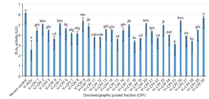
Inhibitory effects of chromatographic pooled fractions (CPF1-CPF30) from the ethyl acetate fraction of Sclerocarya birrea root-bark extract on Naja nigricollis venom PLA2 activity: The venom control displayed the highest activity (6.1±). The CPFs produced varying reductions in enzymatic activity with CPF 25 (3.06±0.05) demonstrated the most significant inhibitory effect. In contrast, fractions such as CPF8, CPF11, CPF20, CPF24, and CPF30 showed comparatively weak inhibition (Fig. 1).
|
|
| Table 4: | GC-MS profile of chromatographic pooled fraction 25 | |||
| Peak no. | Retention time (min) | Total area (%) | Compound name | Molecular formula | Molecular weight (g/mol) |
| 1 | 13.256 | 0.74 | 2-octenal | C12H22O | 126 |
| 2 | 17.071 | 6.28 | Eicosanoic acid | C20H40O2 | 312 |
| 3 | 20.16 | 57.83 | Hexadecanoic acid | C16H32O2 | 256 |
| 4 | 21.074 | 12.46 | Erucic acid | C22H42O2 | 338 |
| 5 | 21.715 | 2.33 | 1,2-Cyclopentanediol | C5H10O2 | 102 |
| 6 | 23.132 | 2.22 | Cyclotetradecane | C14H28 | 196 |
| 7 | 23.586 | 7.85 | 13-octadecenal | C18H34O | 266 |
| 8 | 24.286 | 4.09 | Di-n-octylphthalate | C24H38O4 | 390 |
| 9 | 25.748 | 6.18 | 9,12-Octadecadienoyl chloride | C18H31ClO | 298 |
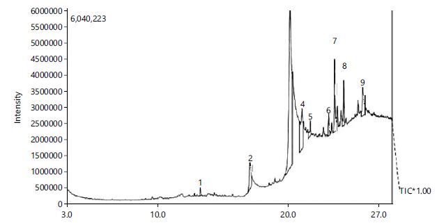
GC-MS profile of chromatographic pooled fraction 25: Figure 2 shows the GC-MS chromatogram of the chromatographic pooled fraction (Fraction 25). A total of nine major compounds were identified in CPF-25. The predominant constituents were hexadecanoic acid (57.83%), erucic acid (12.46%), di-n-octyl phthalate (7.85%), 9,12-octadecadienoyl chloride (6.18%), and 13-octadecenal (4.09%). The identified compounds and their corresponding percentage peak areas are presented in Table 4.
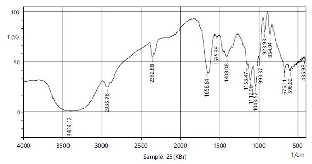
FTIR profile of chromatographic pooled fraction 25: The FTIR spectrum showed characteristic absorption bands at 3381 cm–1 (O-H stretching), 1780 cm–1 (C = O stretching), and 1641 cm–1 (C = C stretching). Additional bands observed between 611.5 to 675.1 cm–1 correspond to C-Cl/C-Br vibrations (Table 5, Fig. 3).
|
|
| Table 5: | FTIR analysis, showing prominent peaks and functional groups in PF 25 | |||
| S/No. | Wavenumber (cm–1) | Transmittance (%) | Bond/Vibration | Functional group |
| 1 | 3381.33 | 0.32 | O-H Stretch | Hydroxyl (alcohol/phenol) |
| 2 | 2933.83 | 12.85 | C-H Stretch | Alkane |
| 3 | 1780.63 | 34.25 | C = O Stretch | Carbonyl (ester/lactone) |
| 4 | 1641.48 | 29.7 | C = C Stretch | Alkene/Aromatic |
| 5 | 1408.08 | 51.02 | O-H Bend | Alcohol |
| 6 | 1153.47 | 39.37 | C-N Stretch | Secondary amine |
| 7 | 1043.37 | 25.5 | C-O Stretch | Alcohol/Ether |
| 8 | 854.49 | 80.95 | C-H Bend | Aromatic/alkene |
| 9 | 675.11 | 41.21 | C-Br Stretch | Halo compound |
| 10 | 611.5 | 44.17 | C-Cl Stretch | Halo compound |
| Table 6: | UV-vis profile of chromatographic pooled fraction 25 | |||
| S/No. | Wavelength (nm) | Absorbance |
| 1 | 310.5 | 0.467 |
| 2 | 280.5 | 0.474 |
| 3 | 268.5 | 0.481 |
| 4 | 259.5 | 0.483 |
| 5 | 242.5 | 0.483 |
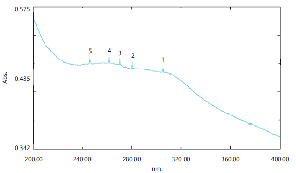
UV-Vis profile of chromatographic pooled fraction 25: The UV-Vis spectrum of CPF25 exhibited absorption maxima between 242.5 and 310.5 nm, indicative of π→π* and n→π* electronic transitions (Table 6, Fig. 4).
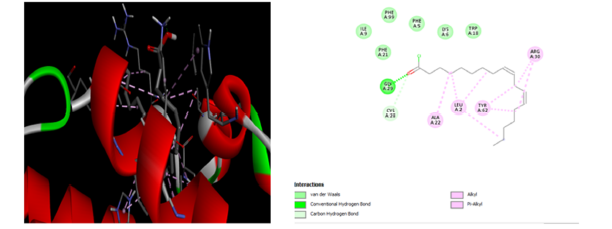
Molecular docking: Docking of 9,12-octadecadienoyl chloride with N. nigricollis PLA2 revealed a binding energy of - 6.29kj/mol. Hydrogen bond were formed with GLY A:29 and CYS A:28; hydrophobic interaction occurred with ALA A:22, LEU A:2 and TYR A:62 as well as vander waals interaction with ILE A:9, PHE A:21, PHE A:99, PHE A:5, lysine (LYS A:6), and TRPA:18 (Fig. 5), indicating active-site occupancy and plausible enzyme inhibition.
DISCUSSION
The present study provides integrated in vivo, in vitro, and in silico evidence supporting the antivenom potential of Sclerocarya birrea root-bark against Naja nigricollis venom. The dose-dependent prolongation of survival observed in envenomed rats treated with the crude methanolic extract indicates a genuine protective effect rather than a nonspecific or symptomatic response. Survival-based bioassays remain a widely accepted and reliable approach for assessing antivenom efficacy and venom neutralization in vivo14. The extract's ability to significantly extend survival time demonstrates its capacity to mitigate the lethal effects of cobra venom. Among the solvent fractions evaluated, the ethyl acetate fraction (EAF) exhibited superior antivenom activity compared with the n-hexane, n-butanol, and aqueous fractions. This observation suggests that the principal bioactive antivenom constituents of S. birrea root-bark are predominantly moderately polar compounds. Similar findings have been reported in natural product-based antivenom studies, where medium-polarity fractions often concentrate enzyme-inhibitory phytochemicals16. The close agreement between in vivo survival outcomes and in vitro phospholipase A2 (PLA2) inhibition further highlights enzyme neutralization as a central mechanism underlying the observed antivenom activity17. PLA2 enzymes are key mediators of cobra venom toxicity, contributing to membrane disruption, inflammation, myonecrosis, and hemolysis by hydrolyzing membrane phospholipids and amplifying inflammatory cascades18. Although conventional antivenoms are effective in neutralizing systemic venom effects, they often show limited efficacy against PLA2-mediated local tissue damage, which can persist even after successful antivenom administration7. The marked PLA2 inhibitory activity exhibited by the EAF therefore provides a mechanistic explanation for its pronounced protective effect in vivo and underscores the therapeutic relevance of targeting venom enzymes directly. Bioactivity-guided chromatographic separation of the EAF led to the identification of chromatographic pooled fraction 25 (CPF-25) as the most potent sub-fraction, achieving over 90% inhibition of venom PLA2 activity. The substantial difference in inhibitory activity between CPF-25 and other pooled fractions confirms the effectiveness of the bioactivity-guided isolation approach in enriching the principal active constituents. Such fractionation strategies are well established in natural product research and are critical for reducing chemical complexity while enhancing biological specificity19,20. Chemical characterization of CPF-25 by GC-MS revealed a predominance of long-chain fatty acid derivatives, including hexadecanoic acid, erucic acid, and 9,12-octadecadienoyl chloride. Compounds belonging to this chemical class have been reported to interact with lipid-processing enzymes and membrane-associated proteins, thereby interfering with phospholipid metabolism and enzyme catalysis6,18. The presence of hydroxyl, carbonyl, and unsaturated functional groups, as confirmed by FTIR and UV-Visible spectroscopy, further supports the capacity of these molecules to participate in hydrogen bonding and hydrophobic interactions with enzymatic active sites. Similar structural features have been associated with effective PLA2 inhibition in other plant-derived antivenom compounds21. Molecular docking analysis provided molecular-level support for the experimental findings by demonstrating stable binding of 9,12-octadecadienoyl chloride within the catalytic pocket of N. nigricollis PLA2. The observed hydrogen bonding and hydrophobic interactions with key amino acid residues indicate effective active-site occupancy and support a plausible competitive or mixed inhibition mechanism. Comparable binding patterns and interaction profiles have been reported for known PLA2 inhibitors, including both synthetic molecules and plant-derived compounds11,22. The favorable binding energy obtained in this study further corroborates the strong in vitro inhibitory activity of CPF-25.
Beyond direct enzyme inhibition, the antivenom efficacy of S. birrea root-bark may also be augmented by ancillary antioxidant and anti-inflammatory effects. Cobra venom-induced tissue injury is often exacerbated by oxidative stress and inflammatory cascades, which contribute to secondary damage at the site of envenomation2,6. Previous studies have demonstrated that S. birrea possesses significant antioxidant and anti-inflammatory properties, which may synergistically complement PLA2 inhibition to limit venom-induced pathology10,23. This multi-target mode of action aligns with the complex pathophysiology of snakebite envenomation and provides a strong scientific basis for the ethnomedicinal use of S. birrea in traditional snakebite management.
Taking together, the findings of this study demonstrate that the antivenom activity of S. birrea root-bark is mediated primarily through direct inhibition of venom PLA2, facilitated by enrichment of lipid-derived bioactive compounds. The integration of in vivo, in vitro, and in silico approaches strengthens the validity of these conclusions and highlights the potential of S. birrea as a source of novel plant-based antivenom adjuncts.
CONCLUSION
This study demonstrates that S. birrea root-bark extract contains bioactive compounds capable of neutralizing N. nigricollis venom, both in vivo and in vitro. The ethyl acetate fraction (EAF) displayed the highest efficacy, and sub-fraction CPF-25 rich in fatty acid derivatives and unsaturated aldehyde, emerged as the most potent inhibitor of PLA2. The structural characterization (GC-MS, FTIR, UV-Vis) and molecular docking of 9,12-Octadecadienoyl chloride substantiate a mechanism of action involving enzyme-active site binding and lipid hydrolysis inhibition. These findings validate the ethno-pharmacological use of S. birrea and underscore its potential as a cost effective plant-based adjuvant for snakebite therapy.
SIGNIFICANCE STATEMENT
Snakebite envenomation caused by Naja nigricollis remains a serious and often overlooked health problem in Sub-Saharan Africa, largely due to limited access to safe and effective antivenom. This study shows that the root-bark of Sclerocarya birrea, a plant widely used in traditional medicine, contains compounds capable of strongly inhibiting venom phospholipase A2, a major enzyme responsible for cobra venom toxicity and tissue damage. Using a bioactivity-guided approach that combines animal studies, enzyme inhibition assays, chemical profiling, and molecular docking, a highly active fraction enriched with fatty acid derivatives that interact favorably with the venom enzyme was identified. These findings provide scientific support for the traditional use of S. birrea and suggest its potential as a plant-based adjunct to conventional antivenom therapy, especially in resource-limited regions.
REFERENCES
- WHO, 2019. Snakebite Envenoming: A Strategy for Prevention and Control. World Health Organization, Geneva, Switzerland, ISBN: 978 92 4 151564 1, Pages: 70.
- Gutiérrez, J.M., J.J. Calvete, A.G. Habib, R.A. Harrison, D.J. Williams and D.A. Warrell, 2017. Snakebite envenoming. Nat. Rev. Dis. Primers, 3.
- Chippaux, J.P., 2017. Snakebite envenomation turns again into a neglected tropical disease!. J. Venomous Anim. Toxins Incl. Trop. Dis., 23.
- Iliyasu, G., F.M. Dayyab, G.C. Michael, M. Hamza, M.A. Habib, J.M. Gutiérrez and A.G. Habib, 2023. Case fatality rate and burden of snakebite envenoming in children-a systematic review and meta-analysis. Toxicon, 234.
- Bittenbinder, M.A., J. van Thiel, F.C. Cardoso, N.R. Casewell, J.M. Gutiérrez, J. Kool and F.J. Vonk, 2024. Tissue damaging toxins in snake venoms: Mechanisms of action, pathophysiology and treatment strategies. Commun. Biol., 7.
- Kini, R.M., 2003. Excitement ahead: Structure, function and mechanism of snake venom phospholipase A2 enzymes. Toxicon, 42: 827-840.
- Uko, S.O., I. Malami, K.G. Ibrahim, N. Lawal and M.B. Bello, M.B. Abubakar and M.U. Imam, 2024. Revolutionizing snakebite care with novel antivenoms: Breakthroughs and barriers. Heliyon, 10.
- Samy, R.P., P. Gopalakrishnakone and V.T.K. Chow, 2012. Therapeutic application of natural inhibitors against snake venom phospholipase A2. Bioinformation, 8: 48-57.
- Ojewole, J.A.O., T. Mawoza, W.D.H. Chiwororo and P.M.O. Owira, 2010. Sclerocarya birrea (A. Rich) Hochst. ['Marula'] (Anacardiaceae): A review of its phytochemistry, pharmacology and toxicology and its ethnomedicinal uses. Phytother. Res., 24: 633-639.
- Konaré, M.A., H.B. Ibrahim, F. Sanogo, C. Cisse, I. Togola and N. Diarra, 2024. Phytochemistry and antioxidant activity of Sclerocarya birrea extracts, A plant used in the traditional management of hypertension in Mali. Int. J. Biosci., 25: 75-82.
- Morris, G.M., R. Huey, W. Lindstrom, M.F. Sanner, R.K. Belew, D.S. Goodsell and A.J. Olson, 2009. AutoDock4 and AutoDockTools4: Automated docking with selective receptor flexibility. J. Comput. Chem., 30: 2785-2791.
- Ma, Z., A. Ajibade and X. Zou, 2024. Docking strategies for predicting protein-ligand interactions and their application to structure-based drug design. Commun. Inf. Syst., 24: 199-230.
- Abubakar, I.S., S.B. Abubakar, A.G. Habib, A. Nasidi and Nandul Durfa et al., 2010. Randomised controlled double-blind non-inferiority trial of two antivenoms for saw-scaled or carpet viper (Echis ocellatus) envenoming in Nigeria. PLoS Negl. Trop. Dis., 4.
- Theakston, R.D.G. and H.A. Reid, 1983. Development of simple standard assay procedures for the characterization of snake venoms. Bull. World Health Organ., 61: 949-956.
- Prabha, N., P. Parthiban, P. Arjun and J. Bhuvana, 2025. Bioactive compounds identification in Abutilon indicum through GC-MS analysis and molecular docking study. Rasayan J. Chem., 18: 641-647.
- UPC, 2023. UniProt: The Universal Protein Knowledgebase in 2023. Nucleic Acids Res., 51: D523-D531.
- Lewin, M.R., R.W. Carter, I.A. Matteo, S.P. Samuel, S. Rao, B.G. Fry and P.E. Bickler, 2022. Varespladib in the treatment of snakebite envenoming: Development history and preclinical evidence supporting advancement to clinical trials in patients bitten by venomous snakes. Toxins, 14.
- Dennis, E.A., J. Cao, Y.H. Hsu, V. Magrioti and G. Kokotos, 2011. Phospholipase A2 enzymes: Physical structure, biological function, disease implication, chemical inhibition, and therapeutic intervention. Chem. Rev., 111: 6130-6185.
- Hostettmann, K., A. Marston, K. Ndjoko and J.L. Wolfender, 2000. The potential of African plants as a source of drugs. Cur. Organic Chem., 4: 973-1010.
- Mors, W.B., M.C. do Nascimento, B.M.R. Pereira and N.A. Pereira, 2000. Plant natural products active against snake bite-the molecular approach. Phytochemistry, 55: 627-642.
- Dev, M. and M. Mukadam, 2025. Functional group profiling of medicinal plants using FTIR spectroscopy. World J. Biol. Pharm. Health Sci., 21: 243-249.
- Kazandjian, T.D., D. Petras, S.D. Robinson, J. van Thiel and H.W. Greene et al., 2021. Convergent evolution of pain-inducing defensive venom components in spitting cobras. Science, 371: 386-390.
- Ojewole, J.A.O., 2003. Hypoglycemic effect of Sclerocarya birrea {(A. Rich.) Hochst.} [Anacardiaceae] stem-bark aqueous extract in rats. Phytomedicine, 10: 675-681.
How to Cite this paper?
APA-7 Style
Sani,
I., Ukwuani-Kwaja,
A.N., Muhammad,
H., Bello,
F., Hassan Fakai,
H.J., Kankara,
A.I., Abdulrauf,
M.S. (2026). Bioactivity-Guided Isolation and Molecular Docking of Potential Anti Naja nigricollis Venom Agent from Sclerocarya birrea Root-Bark Extracts. Trends in Pharmacology and Toxicology, 2(2), 120-129. https://doi.org/10.21124/tpt.2026.120.129
ACS Style
Sani,
I.; Ukwuani-Kwaja,
A.N.; Muhammad,
H.; Bello,
F.; Hassan Fakai,
H.J.; Kankara,
A.I.; Abdulrauf,
M.S. Bioactivity-Guided Isolation and Molecular Docking of Potential Anti Naja nigricollis Venom Agent from Sclerocarya birrea Root-Bark Extracts. Trends Pharm. Toxicol. 2026, 2, 120-129. https://doi.org/10.21124/tpt.2026.120.129
AMA Style
Sani
I, Ukwuani-Kwaja
AN, Muhammad
H, Bello
F, Hassan Fakai
HJ, Kankara
AI, Abdulrauf
MS. Bioactivity-Guided Isolation and Molecular Docking of Potential Anti Naja nigricollis Venom Agent from Sclerocarya birrea Root-Bark Extracts. Trends in Pharmacology and Toxicology. 2026; 2(2): 120-129. https://doi.org/10.21124/tpt.2026.120.129
Chicago/Turabian Style
Sani, Ibrahim, Angela N. Ukwuani-Kwaja, Hasimu Muhammad, Fatima Bello, Habiba Joy Hassan Fakai, Aliyu Idris Kankara, and Muhammad Shafi'u Abdulrauf.
2026. "Bioactivity-Guided Isolation and Molecular Docking of Potential Anti Naja nigricollis Venom Agent from Sclerocarya birrea Root-Bark Extracts" Trends in Pharmacology and Toxicology 2, no. 2: 120-129. https://doi.org/10.21124/tpt.2026.120.129

This work is licensed under a Creative Commons Attribution 4.0 International License.