
Bioinformatics-Guided Analysis of MLH1 Gene Variants: Structural Modeling, Dynamics, and Drug Interaction Studies
-
Amina Islam
Department of Microbiology and Molecular Genetics, University of Okara, 56130, Renala Khurd, Pakistan
Kainat Ramzan
Department of Molecular Biology, University of Okara, 56130, Renala Khurd, Pakistan
Izwa IslamDepartment of Biotechnology, University of Okara, 56130, Renala Khurd, Pakistan
| Received 13 Apr, 2026 |
Accepted 24 Jul, 2026 |
Published 30 Sep, 2026 |
Background and Objective: The MLH1 gene encodes a key protein of the DNA mismatch repair (MMR) system responsible for maintaining genomic stability. Mutations in MLH1 can disrupt DNA repair, leading to microsatellite instability and an increased risk of hereditary cancers such as Lynch syndrome and colorectal cancer. This study aimed to identify deleterious missense variants of the MLH1 gene and evaluate their structural, dynamic, and therapeutic implications using integrated bioinformatics analysis, Normal Mode Analysis (iMODS), and molecular docking studies. Materials and Methods: Missense variants were screened using twelve in silico prediction tools to identify potentially damaging nsSNPs. Mutation3D was used to analyze the spatial clustering of variants. A full-length three-dimensional structure of MLH1 (756 amino acids) was predicted using AlphaFold and validated using QMEAN and ProSA scores. Structural dynamics were examined using iMODs server. Molecular docking was performed in PyRx to assess interactions between wild-type and mutant MLH1 proteins and selected FDA-approved anticancer drugs. Results: Twenty-six nsSNPs were predicted to be highly deleterious, with eight variants located in structurally important regions. Structural validation of the MLH1 model yielded a QMEAN Z-score of -4.17 and a ProSA Z-score of -7.15. Four variants (L11V, R27Q, I115T, and E319K) showed notable structural deviations from the wild-type protein. Normal Mode Analysis indicated that the L11V variant exhibited slightly increased rigidity compared with the wild type, while R27Q, I115T, and E319K showed marginally increased flexibility. Docking analysis revealed stable interactions between MLH1 proteins and several anticancer drugs, including Cabozantinib, Irinotecan, Crizotinib, and Olaparib, although mutations altered binding orientations and interaction patterns. Conclusion: The findings highlight the structural and dynamic effects of MLH1 missense mutations and their potential influence on therapeutic interactions. This study provides insights into MLH1 variant pathogenicity and may support future experimental validation and targeted therapeutic strategies for colorectal cancer.
| Copyright © 2026 Islam et al. This is an open-access article distributed under the Creative Commons Attribution License, which permits unrestricted use, distribution, and reproduction in any medium, provided the original work is properly cited. |
INTRODUCTION
Colorectal cancer (CRC) is a malignant neoplasm of the colon or rectum characterized by uncontrolled cellular proliferation, frequently arising from precancerous polyps. Clinically, CRC manifests with a range of symptoms, including alterations in bowel habits, rectal bleeding, abdominal discomfort, and unintended weight loss1. Globally, CRC represents a significant public health burden, ranking as the third most commonly diagnosed cancer and the 3rd leading cause of cancer-related mortality2. In 2020, more than 1.9 million new CRC cases and over 930,000 deaths were reported3. Notably, early-onset CRC is increasing, particularly in New Zealand, Chile, and England, a trend attributed to unhealthy dietary patterns, physical inactivity, and rising obesity prevalence4. According to the National Cancer Registry of Pakistan, CRC accounted for approximately 4.9% of all cancer cases between 2015 and 2019, ranking the most prevalent cancers in the country5.
Moreover, CRC develops through a multifactorial process involving both genetic susceptibility and environmental influences. Inherited disorders such as Lynch syndrome (LS) and familial adenomatous polyposis (FAP) markedly increase the likelihood of developing CRC by impairing essential DNA repair systems and tumor-suppressing mechanisms. Lynch syndrome, also referred to as hereditary nonpolyposis colorectal cancer (HNPCC), is primarily caused by alterations in DNA mismatch repair (MMR) genes, including MLH1, MSH2, MSH6, and PMS26,7.
In LS, defective MMR leads to the accumulation of DNA replication errors, significantly increasing the CRC cases and other malignancies, such as endometrial and gastric cancers8. To date, more than 3,000 distinct MLH1 variants have been documented, including missense mutations (Leu749Pro, Gly67Arg), splice-site mutations (c.588+5G>A), and frameshift mutations (c.1852_1853delAG). Germline mutations, such as c.677G>A (Val226Ile), destabilize the protein, reduce DNA repair efficiency, and increase the risk of colorectal cancer9.
In sporadic colorectal cancers, MLH1 promoter hypermethylation is a common mechanism of gene silencing, further reducing MMR efficiency and promoting tumorigenesis. In Lynch syndrome, MLH1 mutations account for approximately 25-40% of all identified mismatch repair gene mutations, making it one of the leading genetic contributors to hereditary nonpolyposis colorectal cancer10. The MLH1 gene is located on human chromosome 3p22.2 and spans about 57 kilobases, comprising 19 exons that encode the full-length protein. Exon 1 contains the untranslated region (UTR), while exons 2-19 encode the functional domains of the protein. The MLH1 protein consists of 756 amino acids and forms a heterodimer with PMS2, generating the MutLα complex, which coordinates the repair of base-base mismatches and insertion-deletion loops. The N-terminal ATPase domain (residues 1-340) mediates ATP binding and hydrolysis, providing the energy required for conformational changes during DNA repair. The C-terminal domain (residues 492-756) is responsible for dimerization with PMS2, a critical step for MutLα complex formation and recruitment to the mismatch site11.
Mutations in the ATPase or dimerization domains can severely compromise the protein’s function, resulting in MMR deficiency and genomic instability. MLH1 is associated with germline and somatic alterations, which can disrupt transcription or protein activity12. This study aimed to identify deleterious missense variants of the MLH1 gene using integrated in silico approaches. Functional nsSNPs were evaluated for their disease association, effects on protein stability, and structural flexibility. Three-dimensional modeling and refinement were performed to assess mutation-induced structural changes, while protein dynamics were examined using Normal Mode Analysis through the iMODs server. In addition, potential small-molecule ligands were retrieved from the ZINC and PubChem databases, and protein-ligand interactions were analyzed using molecular docking and Discovery Studio to explore possible therapeutic targets.
MATERIALS AND METHODS
Datasets screening: The reference sequence of the human MLH1 gene was retrieved from the National Center for Biotechnology Information database, and corresponding protein information was obtained from UniProt13. Human SNP data were retrieved from the dbSNP database of the National Center for Biotechnology Information (accessed on 2 October 2025). For downstream analysis, only non-synonymous (missense) SNPs with available protein-level information were selected. These datasets provided the foundation for computational screening and the identification of potentially deleterious variants (Table 1).
SNPs annotation and disease analysis: SNPnexus was used as an integrated platform incorporating multiple prediction tools, including SIFT and PolyPhen. PolyPhen-2, ranging from 0 (benign) to 1 (probably damaging), was used to assess the pathogenicity of variants based on sequence conservation and structural features. PANTHER was employed to analyse evolutionary conservation and Gene Ontology-based functional annotations, supporting SNP pathogenicity prediction. In addition, PredictSNP was used to generate confidence-weighted consensus predictions by integrating multiple algorithms. The potential disease association of the variants was evaluated using PhD-SNP, SNP & GO. SNP & GO combines Gene Ontology annotations with a probability score between 0 and 1; variants with a score>0.5 are considered likely disease-associated variants. SUSPECT was employed to assess the potential impact of these missense mutations on protein stability14,15.
Analysis of MLH1 stability: The impact of nsSNPs on MLH1 protein stability was analysed using multiple computational tools. MuPro predicts stability changes based on machine learning models, where negative ΔΔG values indicate destabilization. The i-Stable applies a meta-prediction approach to assess sequence-based stability alterations. For more rigorous predictions, DUET was employed16.
STRUCTURE-BASED ALGORITHMS
Swiss modeling: The 3D structure of the wild-type (WT) MLH1 protein was generated using the SWISS-MODEL server The FASTA sequence of MLH1 was submitted, templates were selected based on sequence similarity, and the best-fitting models were constructed. The resulting structures were visualized in PyMOL, with site-specific amino acid substitutions incorporated. Atomic-level optimization and minimization of steric clashes were performed using GalaxyRefine13,17.
| Table 1: | Computational workflow and tools employed for variant and structural analysis | |||
| Analysis type | Tool/Database | Primary application |
| Variant data acquisition | NCBI dbSNP | Retrieval of genetic variants |
| UniProt | Protein sequence and functional annotation | |
| Functional impact assessment | SNPnexus | Variant annotation and functional inference |
| PolyPhen-2 | Prediction of structural and functional effects | |
| PANTHER-PSEP | Evolutionary preservation scoring | |
| PredictSNP | Consensus-based variant effect prediction | |
| Disease association prediction | SNP & GO | Gene Ontology-based disease scoring |
| PhD-SNP | Machine learning-based disease classification | |
| SUSPECT | Functional and structural impact evaluation | |
| Protein stability and dynamics | MuPro | ΔΔG-based stability prediction |
| iStable 2.0 | Meta-prediction of stability alterations | |
| DynaMut2 | Stability and flexibility analysis | |
| DUET | Integrated FoldX-mCSM stability estimation | |
| Structural modeling | SWISS-MODEL | Homology-based 3D structure generation |
| Model refinement | GalaxyRefine | Atomic-level structural optimization |
| Structure quality validation | SAVES Server | Stereochemical and interaction validation |
| Model quality evaluation | QMEAN | Global and local quality scoring |
| ProSA-web | Z-score-based structural assessment | |
| Structural comparison | TM-align | Structural alignment and RMSD calculation |
| Mutation clustering analysis | Mutation3D | Identification of mutational hotspots |
| Protein dynamics analysis | iMODS | Normal Mode Analysis for protein flexibility and stability |
| Ligand library preparation | PubChem | Small-molecule compound retrieval |
| Molecular docking | PyRx (AutoDock Vina) | Protein–ligand interaction analysis |
| Interaction visualization | Discovery Studio Visualizer Ligplot+ |
2D/3D interaction profiling |
Template validation: Model quality was evaluated using the SAVESv6.1 server, including PROCHECK, ERRAT, and VERIFY-3D modules. Ramachandran plots generated by PROCHECK assessed torsional angles, with >90% of residues expected in favorable regions; the MLH1 models showed 95.3% of residues in these regions. ERRAT evaluated non-bonded interactions, while VERIFY-3D compared the 3D models against a high-quality protein database for consistency18. Model quality was further assessed using QMEAN, where higher scores indicate greater similarity to experimentally resolved structures. ProSA-web was used to calculate Z-scores, providing an overall quality estimate compared with native proteins14,17.
Structural alteration analysis: Structural alignment of WT and mutant proteins was performed using TM-align. Template Modeling (TM) scores and Root-Mean-Square Deviation (RMSD) values were calculated, with TM scores near 1 indicating high structural similarity, while higher RMSD values reflected significant conformational change15. The spatial distribution of amino acid substitutions in MLH1 was analysed using Mutation3D. This tool allows identification of clustered or hotspot mutations within experimental or modeled protein structures, providing insight into potential effects on folding and function19.
INTERACTION ANALYSIS AND VISUALIZATION
The toxicity profiles of the selected compounds were evaluated using the ProTox-II web server20. Molecular docking studies were conducted using the PyRx platform, which integrates AutoDock and AutoDock Vina algorithms. Protein and ligand structures were converted into PDBQT format, and grid boxes were defined to encompass the active site region. Docking simulations were carried out using the Lamarckian Genetic Algorithm (LGA), and the most favorable binding poses were selected based on minimum binding energy values. The docked protein-ligand complexes were evaluated using Discovery Studio Visualizer and LigPlot+. Interaction profiles were generated in both 2D and 3D formats to identify hydrogen bonds, hydrophobic interactions, and the key amino acid residues contributing to ligand binding within the active site13,14.
NORMAL MODE ANALYSIS OF MLH1 STRUCTURAL DYNAMICS
To evaluate the structural flexibility and dynamic behavior of the protein encoded by the MLH1 gene, normal mode analysis (NMA) was performed using the iMODS server. The analysis was conducted with default parameters. This approach provides insight into protein stability, flexibility, and deformation patterns, allowing assessment of how specific amino acid substitutions may influence the structural behavior of both wild-type and mutant MLH1 proteins18.
RESULTS
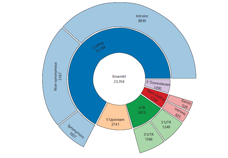
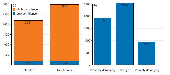
Functional assessment of MLH1 variants: The analysis of the MLH1 gene in the NCBI dbSNP database identified 23,356 SNPs. These included 1,657 synonymous variants and 5,167 missense (non-synonymous) variants. This study found 15,754 were located in coding regions and 1,154 in non-coding regions. Genomic region distribution showed 8,930 intronic SNPs, 529 exonic SNPs, and 2,615 SNPs within untranslated regions, comprising 1,366 variants in the 3-UTR and 1,249 in the 5-UTR. In addition, 1,092 SNPs were identified in the 3 downstream region and 2,741 in the 5 upstream region (Fig. 1). Functional annotation of missense SNPs using SNPnexus and SIFT predicted 2,962 variants as deleterious, 193 as low-confidence deleterious, 2,144 as tolerated, and 161 as low-confidence tolerated (Fig. 2a). PolyPhen analysis classified 1,934 variants as probably damaging, 927 as possibly damaging, and 2,574 as benign (Fig. 2b). Comparative analysis of SIFT and PolyPhen predictions identified 76 common ns SNPs consistently classified as deleterious or probably damaging (Table 2).
|
|
PANTHER analysis predicted three functional outcome categories for the 76 nsSNPs, such as probably damaging, possibly damaging, and benign. Of these, 74 nsSNPs were classified as probably damaging, while I219F and I115T were assigned a score of 0.27 (Table 2). PolyPhen-2 analysis categorized 68 nsSNPs as probably damaging, whereas I513T, S332F, D737V, A31S, L135R, H112R, and V16L were classified as possibly damaging. In addition, A129T (rs63750866) was predicted to be benign by PolyPhen-2 (Table 2). PredictSNP analysis indicated deleterious effects for all 76 nsSNPs on protein function, as summarized in Table 2.
| Table 2: | Computational analysis of high-risk missense variants using diverse functional prediction tools | |||
| SIFT | Polyphen | Panther | Predict SNP | Polyphen 2 | ||||||||
| Variation ID | Variant | AA | Eff | Sc | Eff | Sc | Eff | Sc | Eff | Sc (%) | Eff | Sc |
| rs1193754562 | C/G | R10G | Deleterious | 0 | Probably damaging | 0.961 | Probably damaging | 0.85 | Deleterious | 76 | Probably damaging | 1 |
| rs1224959447 | C/G | P747R | Deleterious | 0 | Probably damaging | 0.961 | Probably damaging | 0.85 | Deleterious | 55 | Probably damaging | 0.997 |
| rs1312172811 | T/G | C672W | Deleterious | 0 | Probably damaging | 0.961 | Probably damaging | 0.89 | Deleterious | 87 | Probably damaging | 1 |
| rs1315572872 | G/A | G54R | Deleterious | 0 | Probably damaging | 0.961 | Probably damaging | 0.89 | Deleterious | 87 | Probably damaging | 1 |
| rs1366752604 | G/T | G6W | Deleterious | 0 | Probably damaging | 0.961 | Probably damaging | 0.74 | Deleterious | 76 | Probably damaging | 1 |
| rs138705565 | G/A | R27Q | Deleterious | 0 | Probably damaging | 0.961 | Probably damaging | 0.89 | Deleterious | 61 | Probably damaging | 1 |
| rs1399177067 | A/G | I94V | Deleterious | 0 | Probably damaging | 0.961 | Probably damaging | 0.89 | Deleterious | 63 | Probably damaging | 0.972 |
| rs1413395234 | T/G | Y625D | Deleterious | 0 | Probably damaging | 0.961 | Probably damaging | 0.89 | Deleterious | 76 | Probably damaging | 1 |
| rs141688321 | T/C | I513T | Deleterious | 0 | Probably damaging | 0.961 | Probably damaging | 0.74 | Deleterious | 74 | Possibly damaging | 0.69 |
| rs1430103195 | T/G | F88V | Deleterious | 0 | Probably damaging | 0.961 | Probably damaging | 0.89 | Deleterious | 75 | Probably damaging | 1 |
| rs1443882824 | C/T | T386I | Deleterious | 0 | Probably damaging | 0.961 | Probably damaging | 0.89 | Deleterious | 55 | Probably damaging | 1 |
| rs147342421 | A/C | Q48H | Deleterious | 0 | Probably damaging | 0.961 | Probably damaging | 0.85 | Deleterious | 60 | Probably damaging | 0.998 |
| rs1482654951 | G/C | D304H | Deleterious | 0 | Probably damaging | 0.961 | Probably damaging | 0.95 | Deleterious | 87 | Probably damaging | 1 |
| rs151119913 | C/T | H315Y | Deleterious | 0 | Probably damaging | 0.961 | Probably damaging | 0.86 | Deleterious | 63 | Probably damaging | 0.999 |
| rs1799977 | A/T | I219F | Deleterious | 0 | Probably damaging | 0.961 | Probably benign | 0.27 | Deleterious | 62 | Probably damaging | 0.967 |
| rs182963667 | T/G | F88L | Deleterious | 0 | Probably damaging | 0.961 | Probably damaging | 0.89 | Deleterious | 75 | Probably damaging | 0.998 |
| rs191257018 | C/T | S332F | Deleterious | 0 | Probably damaging | 0.961 | Probably damaging | 0.74 | Deleterious | 60 | Possibly damaging | 0.544 |
| rs200076893 | T/A | Y126N | Deleterious | 0 | Probably damaging | 0.961 | Probably damaging | 0.89 | Deleterious | 51 | Probably damaging | 0.999 |
| rs267607706 | C/G | N38K | Deleterious | 0 | Probably damaging | 0.961 | Probably damaging | 0.95 | Deleterious | 87 | Probably damaging | 1 |
| rs267607725 | G/A | G98S | Deleterious | 0 | Probably damaging | 0.961 | Probably damaging | 0.95 | Deleterious | 87 | Probably damaging | 0.999 |
| rs267607808 | C/T | P309S | Deleterious | 0 | Probably damaging | 0.961 | Probably damaging | 0.95 | Deleterious | 62 | Probably damaging | 1 |
| rs267607885 | A/T | D737V | Deleterious | 0 | Probably damaging | 0.961 | Probably damaging | 0.85 | Deleterious | 87 | Possibly damaging | 0.942 |
| rs267607886 | A/G | Y684C | Deleterious | 0 | Probably damaging | 0.961 | Probably damaging | 0.89 | Deleterious | 76 | Probably damaging | 1 |
| rs35001569 | A/G | K618E | Deleterious | 0 | Probably damaging | 0.961 | Probably damaging | 0.85 | Deleterious | 65 | Probably damaging | 0.998 |
| rs35045067 | A/G | Y548C | Deleterious | 0 | Probably damaging | 0.961 | Probably damaging | 0.89 | Deleterious | 61 | Probably damaging | 1 |
| rs367654552 | C/G | R18G | Deleterious | 0 | Probably damaging | 0.961 | Probably damaging | 0.86 | Deleterious | 87 | Probably damaging | 1 |
| rs377241633 | T/C | F626L | Deleterious | 0 | Probably damaging | 0.961 | Probably damaging | 0.89 | Deleterious | 60 | Probably damaging | 1 |
| rs41295280 | G/C | G22A | Deleterious | 0 | Probably damaging | 0.961 | Probably damaging | 0.95 | Deleterious | 87 | Probably damaging | 0.994 |
| rs41295284 | T/A | L607H | Deleterious | 0 | Probably damaging | 0.961 | Probably damaging | 0.85 | Deleterious | 72 | Probably damaging | 1 |
| rs550914672 | G/A | E331K | Deleterious | 0 | Probably damaging | 0.961 | Probably damaging | 0.85 | Deleterious | 68 | Probably damaging | 0.983 |
| rs56185292 | C/G | S577W | Deleterious | 0 | Probably damaging | 0.961 | Probably damaging | 0.85 | Deleterious | 61 | Probably damaging | 0.994 |
| rs63750555 | A/T | E23D | Deleterious | 0 | Probably damaging | 0.961 | Probably damaging | 0.85 | Deleterious | 87 | Probably damaging | 0.999 |
| rs63750641 | A/G | K84E | Deleterious | 0 | Probably damaging | 0.961 | Probably damaging | 0.95 | Deleterious | 87 | Probably damaging | 1 |
| rs63750650 | A/G | E268G | Deleterious | 0 | Probably damaging | 0.961 | Probably damaging | 0.57 | Deleterious | 60 | Probably damaging | 0.999 |
| rs63750726 | C/T | P654L | Deleterious | 0 | Probably damaging | 0.961 | Probably damaging | 0.95 | Deleterious | 61 | Probably damaging | 1 |
| rs63750760 | C/T | R385C | Deleterious | 0 | Probably damaging | 0.961 | Probably damaging | 0.89 | Deleterious | 87 | Probably damaging | 1 |
| rs63750792 | C/T | P28L | Deleterious | 0 | Probably Damaging | 0.961 | Probably damaging | 0.89 | Deleterious | 87 | Probably damaging | 1 |
| rs63750796 | G/A | E319K | Deleterious | 0 | Probably damaging | 0.961 | Probably damaging | 0.89 | Deleterious | 61 | Probably damaging | 0.994 |
| rs63750866 | G/A | A120T | Deleterious | 0 | Probably damaging | 0.961 | Probably damaging | 0.85 | Deleterious | 61 | Benign | 0.001 |
| rs63750877 | A/C | K57Q | Deleterious | 0 | Probably damaging | 0.961 | Probably damaging | 0.89 | Deleterious | 63 | Probably damaging | 1 |
| rs63750899 | C/T | P648S | Deleterious | 0 | Probably damaging | 0.961 | Probably damaging | 0.89 | Deleterious | 61 | Probably damaging | 1 |
| rs63750952 | A/G | N64S | Deleterious | 0 | Probably damaging | 0.961 | Probably damaging | 0.89 | Deleterious | 87 | Probably damaging | 1 |
| rs63751109 | C/T | S44F | Deleterious | 0 | Probably damaging | 0.961 | Probably damaging | 0.85 | Deleterious | 87 | Probably damaging | 0.999 |
| rs63751275 | C/T | R687W | Deleterious | 0 | Probably damaging | 0.961 | Probably damaging | 0.74 | Deleterious | 76 | Probably damaging | 1 |
| rs63751597 | C/T | H264Y | Deleterious | 0 | Probably damaging | 0.961 | Probably damaging | 0.85 | Deleterious | 87 | Probably damaging | 0.997 |
| rs63751715 | G/C | Q346H | Deleterious | 0 | Probably damaging | 0.961 | Probably damaging | 0.89 | Deleterious | 72 | Probably damaging | 1 |
| rs730882127 | C/G | A31G | Deleterious | 0 | Probably damaging | 0.961 | Probably damaging | 0.89 | Deleterious | 76 | Probably damaging | 1 |
| rs746206527 | C/A | T310K | Deleterious | 0 | Probably damaging | 0.961 | Probably damaging | 0.89 | Deleterious | 61 | Probably damaging | 1 |
| rs748417604 | G/A | V152M | Deleterious | 0 | Probably damaging | 0.961 | Probably damaging | 0.95 | Deleterious | 76 | Probably damaging | 1 |
| rs749671520 | G/T | A31S | Deleterious | 0 | Probably damaging | 0.961 | Probably damaging | 0.89 | Deleterious | 77 | Possibly damaging | 0.924 |
| rs752850761 | G/T | R79S | Deleterious | 0 | Probably damaging | 0.961 | Probably damaging | 0.89 | Deleterious | 87 | Probably damaging | 1 |
| rs753233578 | T/G | L135R | Deleterious | 0 | Probably damaging | 0.961 | Probably damaging | 0.85 | Deleterious | 51 | Possibly damaging | 0.862 |
| rs756398627 | C/T | R27W | Deleterious | 0 | Probably damaging | 0.961 | Probably damaging | 0.89 | Deleterious | 87 | Probably damaging | 1 |
| rs759287108 | C/G | T45R | Deleterious | 0 | Probably damaging | 0.961 | Probably damaging | 0.89 | Deleterious | 61 | Probably damaging | 1 |
| rs764120517 | T/C | I115T | Deleterious | 0 | Probably damaging | 0.961 | Probably benign | 0.27 | Deleterious | 61 | Probably damaging | 1 |
| rs770276731 | A/G | T96A | Deleterious | 0 | Probably damaging | 0.961 | Probably damaging | 0.95 | Deleterious | 87 | Probably damaging | 1 |
| rs773647920 | A/G | Y97C | Deleterious | 0 | Probably damaging | 0.961 | Probably damaging | 0.86 | Deleterious | 87 | Probably damaging | 1 |
| rs775914775 | A/G | H112R | Deleterious | 0 | Probably damaging | 0.961 | Probably damaging | 0.89 | Deleterious | 74 | Possibly damaging | 0.909 |
| rs776643257 | G/C | V16L | Deleterious | 0 | Probably damaging | 0.961 | Probably damaging | 0.89 | Deleterious | 65 | Possibly damaging | 0.867 |
| rs777054430 | T/C | Y750H | Deleterious | 0 | Probably damaging | 0.961 | Probably damaging | 0.89 | Deleterious | 72 | Probably damaging | 1 |
| rs779581111 | A/G | T310A | Deleterious | 0 | Probably damaging | 0.961 | Probably damaging | 0.89 | Deleterious | 63 | Probably damaging | 0.998 |
| rs780141938 | C/G | I68M | Deleterious | 0 | Probably damaging | 0.961 | Probably damaging | 0.89 | Deleterious | 61 | Probably damaging | 1 |
| rs780232692 | T/C | C142R | Deleterious | 0 | Probably damaging | 0.961 | Probably damaging | 0.89 | Deleterious | 87 | Probably damaging | 1 |
| rs786202396 | G/T | G634V | Deleterious | 0 | Probably damaging | 0.961 | Probably damaging | 0.89 | Deleterious | 72 | Probably damaging | 1 |
| rs786203362 | A/G | R79G | Deleterious | 0 | Probably damaging | 0.961 | Probably damaging | 0.89 | Deleterious | 87 | Probably damaging | 1 |
| rs786203583 | C/A | L741M | Deleterious | 0 | Probably damaging | 0.961 | Probably damaging | 0.85 | Deleterious | 75 | Probably damaging | 0.983 |
| rs786203745 | A/G | T81A | Deleterious | 0 | Probably damaging | 0.961 | Probably damaging | 0.89 | Deleterious | 65 | Probably damaging | 0.996 |
| rs863224637 | T/G | N30K | Deleterious | 0 | Probably damaging | 0.961 | Probably damaging | 0.89 | Deleterious | 87 | Probably damaging | 1 |
| rs863224638 | T/C | V307A | Deleterious | 0 | Probably damaging | 0.961 | Probably damaging | 0.89 | Deleterious | 87 | Probably damaging | 1 |
| rs863224639 | G/T | V314F | Deleterious | 0 | Probably damaging | 0.961 | Probably damaging | 0.89 | Deleterious | 87 | Probably damaging | 1 |
| rs864622396 | T/C | V15A | Deleterious | 0 | Probably damaging | 0.961 | Probably damaging | 0.89 | Deleterious | 87 | Probably damaging | 1 |
| rs864622485 | G/C | E37D | Deleterious | 0 | Probably damaging | 0.961 | Probably damaging | 0.89 | Deleterious | 76 | Probably damaging | 1 |
| rs864622596 | C/G | L11V | Deleterious | 0 | Probably damaging | 0.961 | Probably damaging | 0.89 | Deleterious | 87 | Probably damaging | 1 |
| rs876659860 | A/C | K196T | Deleterious | 0 | Probably damaging | 0.961 | Probably damaging | 0.89 | Deleterious | 61 | Probably damaging | 1 |
| rs876660301 | G/A | V15M | Deleterious | 0 | Probably damaging | 0.961 | Probably damaging | 0.89 | Deleterious | 87 | Probably damaging | 1 |
| rs876660775 | G/T | K311N | Deleterious | 0 | Probably damaging | 0.961 | Probably damaging | 0.95 | Deleterious | 87 | Probably damaging | 1 |
| *Eff: Effect and Sc: Scorz | ||||||||||||
| Table 3: | Prediction of disease-associated nsSNPs using SNPS&GO, SusPect, and PhD-SNP | |||
| SNP and GO | SuSPect | PhD-SNP | |||||
| Variation ID | A.A | Effect | Score | Score | Prediction | Effect | Score (%) |
| rs1193754562 | R10G | Disease | 1 | 54 | Disease | Disease | 73 |
| rs1224959447 | P747R | Disease | 0 | 35 | Disease | Disease | 68 |
| rs1312172811 | C672W | Disease | 7 | 96 | Disease | Disease | 88 |
| rs1315572872 | G54R | Disease | 0 | 57 | Disease | Disease | 88 |
| rs1366752604 | G6W | Neutral | 4 | 52 | Disease | Disease | 77 |
| rs138705565 | R27Q | Disease | 1 | 74 | Disease | Disease | 88 |
| rs1399177067 | I94V | Neutral | 8 | 65 | Disease | Disease | 89 |
| rs1413395234 | Y625D | Neutral | 1 | 93 | Disease | Disease | 88 |
| rs141688321 | I513T | Neutral | 3 | 38 | Disease | Disease | 58 |
| rs1430103195 | F88V | Disease | 2 | 67 | Disease | Disease | 55 |
| rs1443882824 | T386I | Neutral | 1 | 49 | Disease | Disease | 86 |
| rs147342421 | Q48H | Neutral | 7 | 73 | Disease | Disease | 66 |
| rs1482654951 | D304H | Disease | 5 | 97 | Disease | Disease | 86 |
| rs151119913 | H315Y | Neutral | 1 | 45 | Disease | Disease | 66 |
| rs1799977 | I219F | Disease | 3 | 86 | Disease | Disease | 66 |
| rs182963667 | F88L | Neutral | 1 | 70 | Disease | Disease | 66 |
| rs191257018 | S332F | Neutral | 5 | 56 | Disease | Disease | 66 |
| rs200076893 | Y126N | Disease | 6 | 77 | Disease | Disease | 68 |
| rs267607706 | N38K | Disease | 4 | 99 | Disease | Disease | 88 |
| rs267607725 | G98S | Disease | 6 | 96 | Disease | Disease | 82 |
| rs267607808 | P309S | Disease | 4 | 94 | Disease | Disease | 55 |
| rs267607885 | D737V | Neutral | 1 | 50 | Disease | Disease | 82 |
| rs267607886 | Y684C | Disease | 5 | 93 | Disease | Disease | 88 |
| rs35001569 | K618E | Neutral | 3 | 71 | Disease | Disease | 61 |
| rs35045067 | Y548C | Disease | 3 | 88 | Disease | Disease | 82 |
| rs367654552 | R18G | Disease | 1 | 87 | Disease | Disease | 86 |
| rs377241633 | F626L | Disease | 1 | 84 | Disease | Disease | 86 |
| rs41295280 | G22A | Neutral | 1 | 81 | Disease | Disease | 77 |
| rs41295284 | L607H | Disease | 0 | 79 | Disease | Disease | 68 |
| rs550914672 | E331K | Neutral | 6 | 42 | Disease | Disease | 78 |
| rs56185292 | S577W | Disease | 2 | 45 | Disease | Disease | 82 |
| rs63750555 | E23D | Neutral | 1 | 93 | Disease | Disease | 88 |
| rs63750641 | K84E | Disease | 7 | 95 | Disease | Disease | 88 |
| rs63750650 | E268G | Neutral | 1 | 59 | Disease | Disease | 66 |
| rs63750726 | P654L | Disease | 7 | 96 | Disease | Disease | 88 |
| rs63750760 | R385C | Disease | 3 | 63 | Disease | Disease | 86 |
| rs63750792 | P28L | Neutral | 3 | 87 | Disease | Disease | 86 |
| rs63750796 | E319K | Neutral | 4 | 74 | Disease | Disease | 77 |
| rs63750866 | A120T | Neutral | 3 | 30 | Disease | Disease | 59 |
| rs63750877 | K57Q | Neutral | 8 | 55 | Disease | Disease | 58 |
| rs63750899 | P648S | Disease | 5 | 93 | Disease | Disease | 86 |
| rs63750952 | N64S | Neutral | 1 | 86 | Disease | Disease | 86 |
| rs63751109 | S44F | Disease | 0 | 97 | Disease | Disease | 88 |
| rs63751275 | R687W | Neutral | 3 | 13 | Neutral | Disease | 45 |
| rs63751597 | H264Y | Neutral | 0 | 77 | Disease | Disease | 58 |
| rs63751715 | Q346H | Neutral | 2 | 48 | Disease | Disease | 73 |
| rs730882127 | A31G | Neutral | 3 | 96 | Disease | Disease | 58 |
| rs746206527 | T310K | Disease | 4 | 77 | Disease | Disease | 68 |
| rs748417604 | V152M | Disease | 1 | 88 | Disease | Disease | 86 |
| rs749671520 | A31S | Neutral | 1 | 95 | Disease | Disease | 76 |
| rs752850761 | R79S | Disease | 5 | 84 | Disease | Disease | 82 |
| rs753233578 | L135R | Neutral | 4 | 58 | Disease | Disease | 73 |
| rs756398627 | R27W | Disease | 3 | 90 | Disease | Disease | 88 |
| rs759287108 | T45R | Neutral | 2 | 75 | Disease | Disease | 82 |
| rs764120517 | I115T | Disease | 2 | 95 | Disease | Disease | 51 |
| rs770276731 | T96A | Disease | 0 | 91 | Disease | Disease | 73 |
| rs773647920 | Y97C | Disease | 5 | 86 | Disease | Disease | 77 |
| rs775914775 | H112R | Neutral | 7 | 39 | Disease | Disease | 55 |
| rs776643257 | V16L | Neutral | 2 | 89 | Disease | Disease | 86 |
| rs777054430 | Y750H | Disease | 4 | 92 | Disease | Disease | 88 |
| rs779581111 | T310A | Neutral | 6 | 46 | Disease | Disease | 78 |
| rs780141938 | I68M | Neutral | 7 | 77 | Disease | Disease | 59 |
| rs780232692 | C142R | Disease | 2 | 79 | Disease | Disease | 77 |
| rs786202396 | G634V | Disease | 1 | 79 | Disease | Disease | 88 |
| rs786203362 | R79G | Disease | 6 | 90 | Disease | Disease | 86 |
| rs786203583 | L741M | Neutral | 7 | 69 | Disease | Disease | 72 |
| rs786203745 | T81A | Neutral | 6 | 62 | Disease | Disease | 89 |
| rs863224637 | N30K | Neutral | 0 | 78 | Disease | Disease | 86 |
| rs863224638 | V307A | Disease | 2 | 92 | Disease | Disease | 88 |
| rs863224639 | V314F | Disease | 5 | 96 | Disease | Disease | 82 |
| rs864622396 | V15A | Neutral | 4 | 86 | Disease | Disease | 73 |
| rs864622485 | E37D | Neutral | 4 | 73 | Disease | Disease | 88 |
| rs864622596 | L11V | Disease | 0 | 90 | Disease | Disease | 86 |
| rs876659860 | K196T | Disease | 2 | 75 | Disease | Disease | 51 |
| rs876660301 | V15M | Neutral | 5 | 89 | Disease | Disease | 73 |
| rs876660775 | K311N | Disease | 4 | 96 | Disease | Disease | 77 |
| Table 4: | Integrated in silico evaluation of variants affecting protein structural stability | |||
| i-Stable | MUpro | Duet | |||||
| Variation ID | A.A | Effect | Score | Effect | Score | Effect | Score |
| rs1193754562 | R10G | Decrease | 0.775588 | Decrease | -1 | Stabilising | 0.138 |
| rs1224959447 | P747R | Decrease | 0.711756 | Decrease | -1 | Destabilising | -0.144 |
| rs1312172811 | C672W | Decrease | 0.746844 | Decrease | -0.09647 | Destabilising | -1.565 |
| rs1315572872 | G54R | Increase | 0.611656 | Increase | 0.131007 | Destabilising | -1.372 |
| rs1366752604 | G6W | Increase | 0.558945 | Increase | 0.24325 | Destabilising | -1.093 |
| rs138705565 | R27Q | Decrease | 0.645462 | Decrease | -1 | Destabilising | -0.415 |
| rs1399177067 | I94V | Decrease | 0.646576 | Decrease | -0.80259 | Destabilising | -1.214 |
| rs1413395234 | Y625D | Decrease | 0.599322 | Decrease | -0.44977 | Destabilising | -1.469 |
| rs141688321 | I513T | Decrease | 0.671393 | Decrease | -1 | Destabilising | -2.999 |
| rs1430103195 | F88V | Increase | 0.665845 | Increase | 0.300372 | Destabilising | -0.267 |
| rs1443882824 | T386I | Increase | 0.696248 | Increase | 0.039758 | Stabilising | 0.405 |
| rs147342421 | Q48H | Decrease | 0.738391 | Decrease | -0.32425 | Destabilising | -0.478 |
| rs1482654951 | D304H | Decrease | 0.738391 | Decrease | -0.32425 | Destabilising | -0.249 |
| rs151119913 | H315Y | Increase | 0.641638 | Increase | 0.227079 | Stabilising | 1.259 |
| rs1799977 | I219F | Decrease | 0.697505 | Decrease | -1 | Destabilising | -1.087 |
| rs182963667 | F88L | Increase | 0.683275 | Increase | 0.250489 | Destabilising | -0.148 |
| rs191257018 | S332F | Decrease | 0.637513 | Decrease | -0.19562 | Destabilising | -0.448 |
| rs200076893 | Y126N | Decrease | 0.842354 | Decrease | -1 | Destabilising | -1.663 |
| rs267607706 | N38K | Decrease | 0.705164 | Decrease | -0.29669 | Stabilising | 0.079 |
| rs267607725 | G98S | Decrease | 0.834635 | Decrease | -0.87681 | Destabilising | -1.338 |
| rs267607808 | P309S | Decrease | 0.817149 | Decrease | -0.59174 | Destabilising | -0.186 |
| rs267607885 | D737V | Decrease | 0.691426 | Decrease | -0.36223 | Stabilising | 0.254 |
| rs267607886 | Y684C | Decrease | 0.674578 | Decrease | -0.28919 | Destabilising | -0.459 |
| rs35001569 | K618E | Increase | 0.569278 | Increase | 0.229491 | Destabilising | -1.256 |
| rs35045067 | Y548C | Decrease | 0.693891 | Decrease | -1 | Destabilising | -1.812 |
| rs367654552 | R18G | Decrease | 0.692444 | Decrease | -1 | Destabilising | -1.558 |
| rs377241633 | F626L | Decrease | 0.791955 | Decrease | -0.17678 | Destabilising | -0.392 |
| rs41295280 | G22A | Increase | 0.631844 | Increase | 0.429184 | Destabilising | -0.098 |
| rs41295284 | L607H | Decrease | 0.707733 | Decrease | -0.62096 | Destabilising | -1.565 |
| rs550914672 | E331K | Decrease | 0.713918 | Decrease | -0.4841 | Stabilising | 0.008 |
| rs56185292 | S577W | Decrease | 0.555761 | Decrease | -0.11195 | Destabilising | -0.422 |
| rs63750555 | E23D | Increase | 0.631876 | Increase | 0.206299 | Destabilising | -0.722 |
| rs63750641 | K84E | Increase | 0.627028 | Increase | 0.503611 | Stabilising | 0.66 |
| rs63750650 | E268G | Decrease | 0.704084 | Decrease | -1 | Destabilising | -0.705 |
| rs63750726 | P654L | Increase | 0.63126 | Increase | 0.193497 | Stabilising | 0.322 |
| rs63750760 | R385C | Decrease | 0.738606 | Decrease | -0.16808 | Destabilising | -0.436 |
| rs63750792 | P28L | Decrease | 0.738606 | Decrease | -0.16808 | Stabilising | 0.513 |
| rs63750796 | E319K | Decrease | 0.739107 | Decrease | -0.12521 | Destabilising | -0.287 |
| rs63750866 | A120T | Decrease | 0.693477 | Decrease | -0.55338 | Destabilising | -0.042 |
| rs63750877 | K57Q | Decrease | 0.693477 | Decrease | -0.55338 | Destabilising | -0.364 |
| rs63750899 | P648S | Decrease | 0.83609 | Decrease | -1 | Destabilising | -2.312 |
| rs63750952 | N64S | Decrease | 0.67731 | Decrease | -0.02465 | Stabilising | 0.052 |
| rs63751109 | S44F | Decrease | 0.526191 | Increase | 0.361924 | Destabilising | -1.058 |
| rs63751275 | R687W | Decrease | 0.741712 | Decrease | -0.98293 | Destabilising | -0.595 |
| rs63751597 | H264Y | Increase | 0.699031 | Increase | 0.014046 | Stabilising | 1.178 |
| rs63751715 | Q346H | Increase | 0.588122 | Increase | 0.610292 | Stabilising | 0.261 |
| rs730882127 | A31G | Decrease | 0.822508 | Decrease | -0.31982 | Destabilising | -1.949 |
| rs746206527 | T310K | Increase | 0.649609 | Increase | 0.109074 | Destabilising | -1.108 |
| rs748417604 | V152M | Decrease | 0.784881 | Decrease | -0.69358 | Destabilising | -1.587 |
| rs749671520 | A31S | Decrease | 0.8087 | Decrease | -0.59011 | Destabilising | -2.477 |
| rs752850761 | R79S | Decrease | 0.748267 | Decrease | -0.94111 | Destabilising | -0.772 |
| rs753233578 | L135R | Decrease | 0.737886 | Decrease | -1 | Destabilising | -0.677 |
| rs756398627 | R27W | Decrease | 0.712872 | Decrease | -0.60613 | Destabilising | -0.109 |
| rs759287108 | T45R | Increase | 0.755264 | Increase | 0.349333 | Stabilising | 0.205 |
| rs764120517 | I115T | Decrease | 0.673349 | Decrease | -1 | Destabilising | -3.215 |
| rs770276731 | T96A | Increase | 0.507296 | Decrease | -0.82159 | Destabilising | -0.442 |
| rs773647920 | Y97C | Decrease | 0.668154 | Decrease | -0.0317 | Destabilising | -1.549 |
| rs775914775 | H112R | Increase | 0.6027 | Increase | 0.37764 | Destabilising | -0.641 |
| rs776643257 | V16L | Increase | 0.554638 | Increase | 0.351501 | Destabilising | -0.309 |
| rs777054430 | Y750H | Decrease | 0.621652 | Decrease | -1 | Destabilising | -0.763 |
| rs779581111 | T310A | Decrease | 0.703168 | Decrease | -0.1827 | Destabilising | -0.559 |
| rs780141938 | I68M | Decrease | 0.797461 | Decrease | -1 | Destabilising | -0.849 |
| rs780232692 | C142R | Decrease | 0.751534 | Decrease | -0.3283 | Destabilising | -0.007 |
| rs786202396 | G634V | Increase | 0.530842 | Increase | 0.03186 | Destabilising | -1.059 |
| rs786203362 | R79G | Decrease | 0.644622 | Decrease | -1 | Destabilising | -0.714 |
| rs786203583 | L741M | Decrease | 0.761662 | Decrease | -0.15319 | Stabilising | -0.437 |
| rs786203745 | T81A | Decrease | 0.644493 | Decrease | -1 | Destabilising | -0.39 |
| rs863224637 | N30K | Decrease | 0.583433 | Decrease | -0.44567 | Stabilising | 0.002 |
| rs863224638 | V307A | Decrease | 0.66852 | Decrease | -1 | Destabilising | -1.098 |
| rs863224639 | V314F | Decrease | 0.705583 | Decrease | -1 | Destabilising | -1.46 |
| rs864622396 | V15A | Decrease | 0.794747 | Decrease | -0.68189 | Destabilising | -0.602 |
| rs864622485 | E37D | Decrease | 0.714411 | Decrease | -0.32641 | Destabilising | -2.053 |
| rs864622596 | L11V | Decrease | 0.786045 | Decrease | -1 | Destabilising | -0.103 |
| rs876659860 | K196T | Decrease | 0.850422 | Decrease | -0.69323 | Destabilising | -0.989 |
| rs876660301 | V15M | Decrease | 0.850887 | Decrease | -0.2174 | Destabilising | -0.194 |
| rs876660775 | K311N | Decrease | 0.735552 | Decrease | -0.23809 | Destabilising | -0.797 |
Estimation of disease-associated nsSNPs: According to SNP & Go, 42 nsSNPs were predicted as disease-associated, while 35 were classified as neutral, as shown in Table 3. PhD-SNP indicating that 76 nsSNPs are disease-causing, with predicted probabilities ranging from 51% to 89%, as presented in Table 3. Variants such as C672W (rs1312172811), G54R (rs1315572872), R27Q (rs138705565), and Y684C (rs267607886) showed the highest disease probabilities (≥88%), highlighting their potential pathogenic significance. SuSPect predicted that all 77 nsSNPs are disease-associated, with scores ranging from 13 to 99. The algorithm indicated a high likelihood of functional impact for most variants, with particularly high scores observed for C672W (rs1312172811, score 96), N38K (rs267607706, score 99), and P654L (rs63750726, score 96). Only R687W (rs63751275) showed a low score of 13 and was classified as neutral, as shown in Table 3.
|
| Table 5: | Functional prediction and visualization of the MLH1 gene by mutation 3D | |||
| rs ID | Model | Prediction | rs ID | Model | Prediction |
| rs138705565 | R27Q | Clustered mutation | rs876660775 | K311N | Covered mutation |
| rs1399177067 | I94V | Covered mutation | rs752850761 | R79S | Covered mutation |
| rs147342421 | Q48H | Covered mutation | rs756398627 | R27W | Clustered mutation |
| rs1482654951 | D304H | Covered mutation | rs764120517 | I115T | Covered mutation |
| rs1799977 | I219F | Covered mutation | rs779581111 | T310A | Covered mutation |
| rs200076893 | Y126N | Covered mutation | rs780141938 | I68M | Covered mutation |
| rs267607706 | N38K | Covered mutation | rs780232692 | C142R | Covered mutation |
| rs267607725 | G98S | Covered mutation | rs786203362 | R79G | Clustered mutation |
| rs367654552 | R18G | Covered mutation | rs864622396 | V15A | Clustered mutation |
| rs63750650 | E268G | Covered mutation | rs864622596 | L11V | Clustered mutation |
| rs63750796 | E319K | Covered mutation | rs876659860 | K196T | Covered mutation |
| rs63750877 | K57Q | Clustered mutation | rs876660301 | V15M | Clustered mutation |
| rs63751109 | S44F | Covered mutation | rs730882127 | A31G | Clustered mutation |
Evaluation of protein stability: We assessed the impact of nsSNPs on protein stability using three computational tools. The i-Stable analysis indicated that 19 nsSNPs could increase the stability of the mutant protein, while 57 nsSNPs were predicted to reduce stability, which may negatively affect protein function (Table 4). MUpro results were similar, showing 57 nsSNPs as destabilizing and 19 as stabilizing (Table 4). DUET identified 61 nsSNPs as destabilizing and 15 as stabilizing, demonstrating some differences between the tools (Table 4). By comparing all prediction tools, we identified 26 nsSNPs as highly deleterious, which were chosen for further detailed analysis.
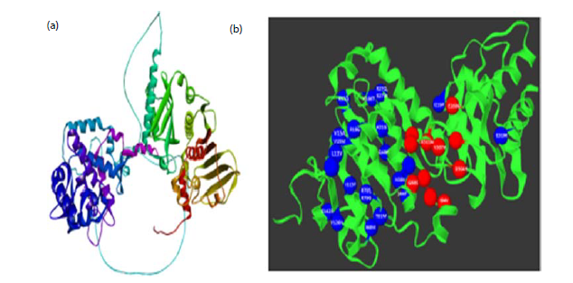
Prediction of mutation clusters: The MLH1 protein was analysed using the Mutation3D web server with all 26 selected nsSNPs as input. Among these, eight missense mutations formed clusters, while the remaining 18 were classified as covered mutations (Table 5). For structural analysis, a 3D model of MLH1 was generated using homology modeling, based on 23 templates obtained from the RCSB PDB database (Fig. 3a). These models, however, did not span the full length of the protein, which consists of 756 amino acids (UniProt ID: P40692). The clustered mutations were R27Q, K57Q, A31G, R27W, R79G, V15A, L11V, and V15M, whereas the covered mutations included I94V, Q48H, D304H, I219F, Y126N, N38K, G98S, R18G, E268G, E319K, S44F, R79S, I115T, T310A, I68M, C142R, K196T, and K311N (Fig. 3b).
SWISS modeling of MLH1 protein: Structural modeling of the human MLH1 protein was performed to evaluate the impact of selected missense variants on protein conformation. A total of 26 non-synonymous substitutions (R27Q, I94V, Q48H, D304H, I219F, Y126N, N38K, G98S, R18G, E268G, E319K, K57Q, S44F, A31G, R79S, R27W, I115T, T310A, I68M, C142R, R79G, V15A, L11V, K196T, V15M, and K311N) were included for comparative homology modeling. Fifty potential templates were identified, among which P40692 showed the highest suitability. The three-dimensional structure of MLH1 was generated using the AlphaFold database model for MLH1_HUMAN (Homo sapiens), based on template P40692.1, covering residues 1-756 with complete sequence coverage. The selected template exhibited 97.98% sequence identity with the query protein. The predicted oligomeric state of the modeled structure was monomeric, and the final model was saved in PDB format for validation.
|
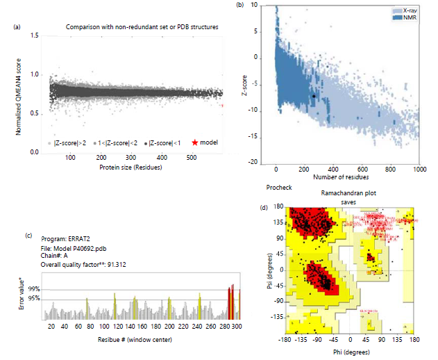
Moreover, Model 1 (P40692.1) demonstrated higher structural quality and was selected for downstream analyses. Model validation using the SAVES server yielded an ERRAT score of 91.312%, indicating reliable non-bonded atomic interactions. Ramachandran plot analysis performed through SWISS-MODEL showed that 90.30% of residues were located in the most favored regions. Additional stereochemical assessment using PROCHECK further confirmed the structural reliability of the model, with detailed statistics summarized in Table 6. The selected structure was refined using GalaxyRefine to improve side-chain conformations and overall structural geometry. Model quality evaluation resulted in a QMEAN Z-score of -4.17, suggesting reduced structural stability (Fig. 4a), while ProSA analysis produced a Z-score of -7.15, supporting the presence of structural instability (Fig. 4b). The optimized P40692.1. A model is highlighted in Fig. 4c and the Procheck Ramachandran Plot (Fig. 4d). Root-Mean-Square Deviation (RMSD) analysis identified L11V (0.57 Å), R27Q (0.57 Å), I115T (0.58 Å), and E319K (0.57 Å), as exhibiting the highest conformational deviations from the wild-type structure. These variants were therefore selected for subsequent protein-ligand docking studies (Table 6).
| Table 6: | Structural validation and comparative assessment of wild-type and mutant MLH1 protein models | |||
| ERRAT | Precheck-ramachandran plot | Verify | TM align | ||||||
| Protein | A.A | Score | Core (%) | Allow (%) | Generously (%) | Disallowed (%) | Score (%) | TM Score | RMSD |
| rs138705565 | R27Q | 93.0116 | 95.1 | 4.1 | 0.6 | 0.1 | 66.4 | 0.99644 | 0.57 |
| rs1399177067 | I94V | 94.5364 | 96.2 | 3.1 | 0.3 | 0.4 | 67.06 | 0.99652 | 0.56 |
| rs147342421 | Q48H | 93.3665 | 95.1 | 4.3 | 0.3 | 0.3 | 66.8 | 0.99651 | 0.56 |
| rs1482654951 | D304H | 93.6982 | 95.4 | 3.7 | 0.6 | 0.3 | 64.55 | 0.99668 | 0.55 |
| rs1799977 | I219F | 93.75 | 94.9 | 4.6 | 0.1 | 0.4 | 66.67 | 0.99652 | 0.56 |
| rs200076893 | Y126N | 94.6755 | 95.9 | 3.5 | 0.1 | 0.4 | 66.27 | 0.99668 | 0.55 |
| rs267607706 | N38K | 94.7811 | 96 | 3.4 | 0.3 | 0.3 | 66.67 | 0.99649 | 0.56 |
| rs267607725 | G98S | 91.6667 | 95.2 | 4.1 | 0.4 | 0.3 | 65.34 | 0.99662 | 0.55 |
| rs367654552 | R18G | 94.5364 | 95.6 | 3.8 | 0.3 | 0.3 | 63.89 | 0.99663 | 0.55 |
| rs63750650 | E268G | 94.3709 | 95.7 | 3.8 | 0.3 | 0.1 | 61.64 | 0.99659 | 0.56 |
| rs63750796 | E319K | 95.0413 | 96 | 3.4 | 0.3 | 0.3 | 65.08 | 0.9965 | 0.57 |
| rs63750877 | K57Q | 93.0693 | 96 | 3.2 | 0.6 | 0.1 | 64.55 | 0.99667 | 0.55 |
| rs63751109 | S44F | 95.1505 | 95.7 | 3.7 | 0.3 | 0.3 | 61.77 | 0.99654 | 0.56 |
| rs730882127 | A31G | 96.8333 | 96 | 3.4 | 0.1 | 0.4 | 64.29 | 0.99667 | 0.55 |
| rs752850761 | R79S | 90.9091 | 95.7 | 3.7 | 0.1 | 0.4 | 67.86 | 0.99672 | 0.55 |
| rs756398627 | R27W | 94.3615 | 95.6 | 3.7 | 0.3 | 0.4 | 63.62 | 0.99658 | 0.56 |
| rs764120517 | I115T | 94.5455 | 95.6 | 3.8 | 0.3 | 0.3 | 67.06 | 0.99635 | 0.58 |
| rs779581111 | T310A | 94.2339 | 95.6 | 3.7 | 0.3 | 0.4 | 66.67 | 0.99661 | 0.55 |
| rs780141938 | I68M | 92.7393 | 96.2 | 3.4 | 0.3 | 0.1 | 66.01 | 0.99657 | 0.56 |
| rs780232692 | C142R | 95.0331 | 96.2 | 3.4 | 0.3 | 0.1 | 62.43 | 0.9966 | 0.56 |
| rs786203362 | R79G | 94.3615 | 95.9 | 3.5 | 0.3 | 0.3 | 67.59 | 0.99663 | 0.55 |
| rs864622396 | V15A | 93.3555 | 95.9 | 3.4 | 0.3 | 0.4 | 66.67 | 0.99658 | 0.56 |
| rs864622596 | L11V | 93.1894 | 95.3 | 4.1 | 0.3 | 0.3 | 68.65 | 0.99644 | 0.57 |
| rs876659860 | K196T | 93.75 | 95.6 | 4.1 | 0.1 | 0.1 | 63.76 | 0.99657 | 0.56 |
| rs876660301 | V15M | 94.1176 | 95.7 | 3.7 | 0.4 | 0.1 | 66.93 | 0.99665 | 0.55 |
| rs876660775 | K311N | 95.5224 | 95.9 | 3.4 | 0.3 | 0.4 | 69.18 | 0.99661 | 0.55 |
| Table 7: | Docking score for 30 ligands with wild-type and mutant MLH1 proteins | |||
| Ligands | Wild type (P40692) | R27Q | E319K | I115T | L11V |
| 5Fluorouracil | -4.6 | -5 | -4.6 | -4.8 | -4.4 |
| Adagrasib | -7.8 | -7.5 | -7.5 | -7.3 | -7.7 |
| Afatinib | -7.4 | -7.5 | -7.6 | -7.6 | -7.4 |
| Bevacizumab | -5.2 | -5.4 | -5.3 | -5.3 | -5.4 |
| Cabozantinib | -8.8 | -9 | -9.1 | -8.2 | -8.8 |
| Camptosar | -8.6 | -8.6 | -9.1 | -8.9 | -8.8 |
| Capecitabine | -6.4 | -6.3 | -6 | -6.6 | -6.3 |
| Crizotinib | -8.5 | -8.4 | -4.2 | -7.6 | -8.2 |
| Doxorubicin | -7.2 | -7.6 | -8.6 | -8.3 | -7.6 |
| Erlotinib | -7.3 | -7 | -7 | -6.8 | -6.7 |
| Fruquintinib | -7.6 | -7.4 | -7.8 | -7.3 | -8 |
| Hydroxyurea | -3.9 | -3.9 | -3.9 | -3.9 | -3.9 |
| Irinotecan | -7.7 | -7.3 | -7.3 | -7.7 | -7.9 |
| Lenvatinib3D | -7.7 | -7.1 | -7.4 | -7.7 | -7.1 |
| Leucovorin | -7.3 | -7.6 | -7.5 | -7.5 | -7.4 |
| Methotrexate | -7.5 | -7.4 | -7.5 | -7.5 | -7.8 |
| Mitomycin | -6.5 | -6.4 | -6.5 | -7.2 | -6.4 |
| Nintedanib | -7.5 | -8.7 | -8.6 | -8.6 | -8.7 |
| Niraparib | -7.6 | -7.6 | -8.1 | -8 | -7.5 |
| Olaparib | -8.5 | -8.3 | -8 | -9.1 | -7.6 |
| Osimertinib | -8.3 | -7.7 | -6.9 | -7.8 | -8.1 |
| Panobinostat | -6.6 | -6.3 | -8.8 | -6.3 | -6.4 |
| Regorafenib | -8 | -8.3 | -7.8 | -8.5 | -7.8 |
| Rucaparib | -8 | -6.8 | -7.5 | -6.9 | -8 |
| Selpercatinib | -8 | -7.9 | -8.7 | -7.7 | -8.2 |
| Sorafenib | -8.3 | -8.1 | -8.1 | -8.3 | -8.4 |
| Sunitinib | -7.3 | -7.3 | -7.3 | -6.8 | -7 |
| Tegafur | -5.9 | -6 | -5.9 | -5.4 | -5.9 |
| Temozolomide | -6.2 | -6.3 | -6.1 | -6 | -5.9 |
| Tivozanib | -7.7 | -7.6 | -7.6 | -7.9 | -7.7 |
| Table 8: | Toxicity prediction of selected anticancer ligands using the ProTox-II server | |||
| Ligands | Carcinogenicity | Immunotoxicity | Mutagenicity | Cytotoxicity | BBB Barrier | Ecotoxicity | Clinical toxicity |
Nutritional toxicity |
| Cabozantinib | Active (0.52) | Active (0.89) | Inactive (0.59) | Inactive (0.66) | Active (0.56) | Active (0.55) | Active (0.75) | Inactive (0.70) |
| Camptosar | Inactive (0.62) | Active (0.96) | Inactive (0.97) | Inactive (0.93) | Inactive (1.00) | Active (0.73) | Inactive (0.56) | Inactive (0.74) |
| Crizotinib | Active (0.55) | Active (0.65) | Inactive (0.56) | Inactive (0.64) | Active (0.67) | Active (0.54) | Active (0.70) | Inactive (0.70) |
| Nintedanib | Inactive (0.52) | Inactive (0.79) | Inactive (0.63) | Inactive (0.56) | Inactive (0.71) | Inactive (0.68) | Active (0.67) | Inactive (0.58) |
| Olaparib | Inactive (0.57) | Inactive (0.95) | Inactive (0.54) | Inactive (0.65) | Active (0.85) | Inactive (0.52) | Active (0.71) | Inactive (0.71) |
| Sorafenib | Inactive (0.50) | Active (0.92) | Inactive (0.79) | Active (0.77) | Active (0.65) | Active (0.50) | Active (0.69) | Inactive (0.70) |
| Regorafenib | Inactive (0.50) | Active (0.99) | Inactive (0.79) | Active (0.77) | Active (0.65) | Active (0.50) | Active (0.69) | Inactive (0.70) |
| Panobinostat | Active (0.57) | Inactive (0.72) | Active (0.55) | Inactive (0.63) | Active (0.78) | Inactive (0.55) | Active (0.87) | Inactive (0.55) |
| Selpercatinib | Inactive (0.57) | Active (0.97) | Inactive (0.58) | Inactive (0.61) | Active (0.59) | Active (0.56) | Active (0.57) | Inactive (0.72) |
In silico protein-ligand docking study: Molecular docking studies were performed using the PyRx platform to investigate the binding interactions between the MLH1 protein and a library of small-molecule compounds. A total of 30 ligands were screened against the MLH1 structure, with detailed compound information presented in Table 7. The selected compounds are well-known or emerging anticancer agents with reported relevance in colorectal cancer therapy. These results highlight the potential therapeutic significance of these molecules in MLH1-mutated or mismatch repair-deficient colorectal cancer and support their further investigation through molecular dynamics simulations and experimental validation.
The docking analysis revealed that Cabozantinib, Camptosar, Crizotinib, Nintedanib, Olaparib, Sorafenib, Regorafenib, Panobinostat, and Selpercatinib exhibited strong binding affinities toward both wild-type and mutant MLH1 proteins. Toxicity prediction using the ProTox-II server indicated generally low mutagenic and nutritional toxicity profiles, although variations were observed in immunotoxic, carcinogenic, cytotoxic, and clinical toxicity endpoints. Among the screened compounds, Nintedanib and Olaparib demonstrated comparatively favorable toxicity profiles, as given in Table 8.
These ligands were further evaluated against MLH1 wild-type and mutant variants (R27Q, L11V, I115T, and E319K), which were identified through RMSD-based structural deviation analysis. Interaction analyses were performed to assess mutation-induced changes in binding affinity, residue contacts, and complex stability. All compounds showed binding energies below -3.9 kcal/mol, indicating stable interactions with both native and mutant MLH1 structures. Notably, Cabozantinib exhibited the highest binding affinity (up to -9.1 kcal/mol), followed by Camptosar and Olaparib, which consistently showed strong docking performance across all protein variants. Furthermore, Nintedanib, Sorafenib, Regorafenib, Panobinostat, and Selpercatinib maintained stable and robust binding interactions with both wild-type and mutant MLH1, supporting their potential as promising inhibitory candidate.
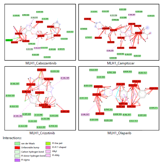
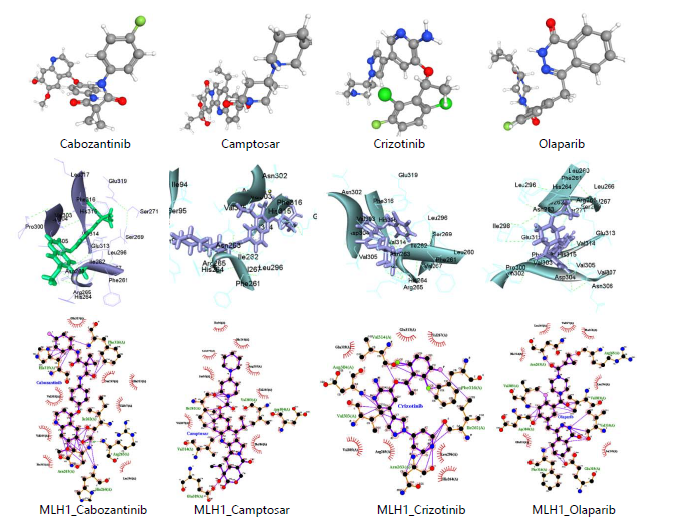
As summarized in Table 9, Cabozantinib established extensive van der Waals interactions with key MLH1 residues, including PRO300, SER95, ARG265, PHE261, LEU260, LEU296, VAL303, ASP304, ILE298, GLU297, GLU313, SER269, SER271, LEU317, and GLU319. Similarly, Camptosar engaged with THR96, ILE94, TYR97, GLU297, LEU296, PHE261, ASN306, LEU260, ILE298, ASN302, SER269, VAL307, LEU317, and SER271. Crizotinib was observed to interact with VAL307, GLU319, GLU313, SER269, GLU268, ILE298, LEU260, ASN302, LEU296, GLU297, HIS264, and PHE261, while Olaparib occupied the binding pocket through contacts with GLU297, LEU296, HIS264, ILE298, LEU266, PRO300, PHE261, LEU260, ASN306, ASN302, VAL307, LEU317, GLU313, SER269, and SER271, as illustrated in Fig. 5. As shown in Fig. 6, Cabozantinib, Camptosar, Crizotinib, and Olaparib demonstrated stable and well-oriented binding within the MLH1 active site. The corresponding docking conformations and LigPlot+ interaction profiles revealed multiple hydrogen bonding and hydrophobic interactions with surrounding residues, indicating strong and energetically favorable protein-ligand complexes.
|
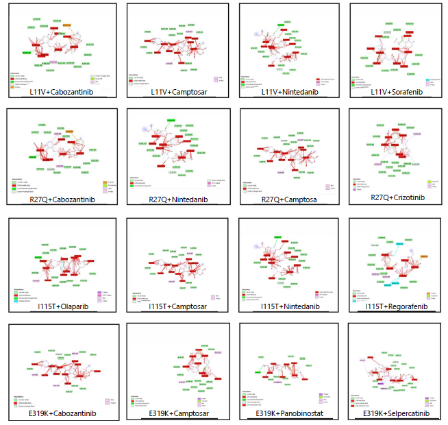
For the L11V mutant, Cabozantinib interacted with ILE298, ARG265, SER95, ILE94, PRO300, ASP304, LEU296, PHE261, GLU297, GLU313, SER269, LEU272, SER271, LEU323, and LEU317. Camptosar formed interactions with ILE94, GLU297, LEU296, PHE261, ILE298, SER269, LEU260, SER271, LEU317, ASN302, VAL307, THR96, and TYR97. Nintedanib engaged VAL307, ASP304, GLU313, ARG265, ILE94, PRO300, SER95, GLU297, GLU268, LEU260, HIS312, ILE298, LEU296, GLU319, and SER271. Sorafenib interacted with GLU297, LEU296, ILE298, VAL305, ASN306, LEU317, VAL307, THR270, SER269, SER271, LEU272, and GLU313 as shown in Fig. 7.
In the R27Q mutant, Cabozantinib formed interactions with SER95, ARG265, ILE94, PHE261, PRO300, LEU296, ILE298, GLU297, ASP304, GLU313, SER269, SER271, LEU272, LEU317, and LEU323, while Camptosar bound with THR96, TYR97, ILE94, GLU297, LEU296, PHE261, LEU260, ILE298, SER269, LEU317, ASN302, GLU313, GLU319, VAL307, and SER271. Crizotinib interacted with GLU319, LEU317, VAL307, GLU313, SER269, GLU268, LEU260, ILE298, GLU297, LEU272, ASN302, PRO300, LEU296, HIS264, PHE261, and ARG265. Nintedanib engaged VAL307, ASP304, GLU313, ILE94, ARG265, PRO300, SER95, GLU297, GLU268, HIS312, LEU260, ILE298, SER269, LEU296, and GLU319as shown in Table 9.
In the I115T mutant, Camptosar interacted with ILE94, THR96, TYR97, GLU297, LEU296, PHE261, ILE298, LEU260, SER269, SER271, VAL307, ASN302, and LEU317. Nintedanib formed van der Waals interactions with VAL307, ASP304, GLU319, LEU296, ILE298, LEU260, HIS312, GLU313, GLU268, GLU297, ARG265, ILE94, SER95, and PRO300. Olaparib interacted through HIS264, LEU296, GLU297, ILE298, PRO300, ASN302, LEU317, LEU266, PHE261, LEU260, ASN306, VAL307, GLU313, SER269, and SER271. Regorafenib engaged LEU317, SER95, THR96, HIS264, GLU297, LEU296, VAL305, ILE298, VAL267, VAL307, and SER269, as shown in Fig. 7.
|
| Table 9: | Interacting residues obtained from docking studies. Variants of the wild protein, R27Q, L11V, I115T and E319K as well as the ligands' hydrophobic interactions and binding residues | |||
| Protein-ligands | Hydrophilic interactions | Hydrophobic interactions | Stearic hinderance |
| WT-Cabozantinib | PRO300, SER95, ARG265, PHE261, | VAL267 | VAL262, ASN263, HIS264, HIS315, |
| LEU260, LEU296, VAL303, ASP304, | VAL305, VAL314, PHE316 | ||
| ILE298, GLU297, GLU313, SER269, | |||
| SER271, LEU317, GLU319 | |||
| WT-Camptosar | THR96, ILE94, TYR97, GLU297, | VAL305, VAL267 | SER95, HIS264, ASN263, ILE262, |
| LEU296, PHE261, ASN306, LEU260, | ARG265, VAL303, ASP304, VAL314, | ||
| ILE298, ASN302, SER269, VAL307, | GLU319, HIS315, PHE316 | ||
| LEU317, SER271 | |||
| WT-Crizotinib | VAL307, GLU319, GLU313, SER269, | VAL305, VAL267 | VAL314, HIS315, ASP304, PHE316, |
| GLU268, ILE298, LEU260, ASN302, | VAL303, ASN263, ILE262, ARG265 | ||
| LEU296, GLU297, HIS264, PHE261 | |||
| WT-Olaparib | GLU297, LEU296, HIS264, ILE298, | VAL267 | ASN263, ILE262, ARG265, VAL303, |
| LEU266, PRO300, PHE261, LEU260, | ASP303, VAL305, PHE316, HIS315, | ||
| ASN306, ASN302, VAL307, LEU317, | VAL314, GLU319 | ||
| GLU313, SER269, SER271 | |||
| R27Q-Cabozantinib | SER95, ARG265, ILE94, PHE261, | VAL303, VAL267, | ASN263, ILE262, HIS264, VAL305, |
| PRO300, LEU296, ILE298, GLU297, | GLU319 | VAL314, HIS315, PHE316 | |
| ASP304, GLU313, SER269, SER271, | |||
| LEU272, LEU317, LEU323 | |||
| R27Q-Camptosar | THR96, TYR97, ILE94, GLU297, | VAL305, VAL267 | SER95, HIS264, ASN263, ARG265, |
| LEU296, PHE261, LEU260 ILE298, | ILE262, VAL303, ASP304, VAL314, | ||
| SER269, LEU317, ASN302, GLU313, | PHE316, HIS315 | ||
| GLU319, VAL307, SER271 | |||
| R27Q-Crizotinib | GLU319, LEU317, VAL307, GLU313, | VAL267, VAL305 | ASP304, HIS315, VAL303, VAL314, |
| SER269, GLU268, LEU260, ILE298, | PHE316, ASN263, ILE262 | ||
| GLU297, LEU272, ASN302, PRO300, | |||
| LEU296, HIS264, PHE261, ARG265 | |||
| R27Q-Nintedanib | VAL307, ASP304, GLU313, ILE94, | NULL | HIS315, PHE316, VAL314, VAL303, |
| ARG265, PRO300, SER95, GLU297, | VAL305, ILE262, VAL267, HIS264, | ||
| GLU268, HIS312, LEU260, ILE298, | ASN263 | ||
| SER269, LEU296, GLU319 | |||
| L11V- Cabozantinib | ILE298, ARG265, SER95, ILE94, | VAL267, GLU319 | VAL305, VAL303, ASN263, HIS264, |
| PRO300, ASP304, LEU296, PHE261, | ILE262, VAL314, HIS315, PHE316 | ||
| GLU297, GLU313, SER269, LEU272, | |||
| SER271, LEU323, LEU317 | |||
| L11V-Camptosar | ILE94, GLU297, LEU296, PHE261, | VAL305, VAL267 | SER95, ASN263, ILE262, ARG265, |
| ILE298, SER269, LEU260, SER271, | HIS264, VAL303, ASP304, VAL314, | ||
| LEU317, ASN302, VAL307, THR96, TYR97 | GLU319, PHE316, HIS315 | ||
| L11V-Nintedanib | VAL307, ASP304, GLU313, ARG265, | NULL | HIS315, PHE316, VAL314, VAL303, |
| ILE94, PRO300, SER95, GLU297, | SER269, ILE262, VAL267, HIS264, | ||
| GLU268, LEU260, HIS312, ILE298, | ASN263, VAL305 | ||
| LEU296, GLU319, SER271 | |||
| L11V-Sorafenib | GLU297, LEU296, ILE298, VAL305, | NULL | HIS264, ASN263, ILE262, ARG265, |
| ASN306, LEU317, VAL307, THR270, | PHE316, VAL303, ASP304, HIS315, | ||
| SER269, SER271, LEU272, GLU313 | VAL314, GLU319 | ||
| I115T-Camptosar | ILE94, THR96, TYR97, GLU297, | VAL305, VAL267 | SER95, HIS264, ASN263, ILE262, |
| LEU296, PHE261, ILE298, LEU260, | ARG265,VAL303, ASP304, VAL314, | ||
| SER269, SER271, VAL307, ASN302,LEU317 | PHE316, GLU319, HIS315, PHE316 | ||
| I115T-Nintedanib | VAL307, ASP304, GLU319, LEU296, | NULL | HIS315, PHE316, VAL314, VAL303, |
| ILE298, LEU260, HIS312, GLU313, | VAL305, SER269, ILE262, VAL267, | ||
| GLU268, GLU297, ARG265, ILE94, | HIS264, ASN263 | ||
| SER95, PRO300 | |||
| I115T-Olaparib | HIS264, LEU296, GLU297, ILE298, | VAL267 | ASN263, ILE262, ARG265, VAL303, |
| PRO300, ASN302, LEU317, LEU266, | ASP304, VAL305, HIS315, PHE316, | ||
| PHE261, LEU260, ASN306, VAL307, | VAL314, GLU319 | ||
| GLU313, SER269, SER271 | |||
| I115T-Regorafenib | LEU317, SER95, THR96, HIS264, | ILE94, ARG265, VAL303, | ASN263, ILE262, PHE316, VAL314, |
| GLU297, LEU296, VAL305, ILE298, | GLU313, ASP304, GLU319 | HIS315 | |
| VAL267, VAL307. SER269 | |||
| E319K-Cabozantinib | THR96, ILE94, GLU297, | VAL267, TYR97, VAL305 | SER95, HIS264, ASN263, ARG265, |
| LEU296, PHE261, ILE298, | ILE262, VAL303, ASP304, VAL314, | ||
| SER269, SER271, LEU317, | PHE316, HIS315, LYS319 | ||
| ASN302, VAL307 | |||
| E319K-Crizotinib | ASN302, PRO300, VAL307, | VAL305, VAL267 | ASP304, HIS315, VAL303, PHE316, |
| LYS319, GLU313, SER269, | VAL314, ASN263, ILE262, ARG265 | ||
| ILE298, HIS264, LEU296, | |||
| LEU272, GLU297, GLU268, PHE261 | |||
| E319K-Panobinostat | ILE94, SER95, PHE261, VAL305, | ARG265, VAL267 | ILE262, ASN263, HIS264, VAL303, |
| ASP304, VAL307, GLU313, SER269, SER271 | VAL314, PHE316, HIS315, LYS319 | ||
| E319K-Selpercatinib | SER271, THR270, LEU272, SER269, | LEU317, VAL303, | LYS319, HIS315, VAL314, PHE316, |
| GLU313, TYR97, VAL305, THR96, | ARG265, VAL267 | ILE262, ASN262, ASN263, ILE94 | |
| ASP304, VAL307, ILE298, GLU297, | |||
| LEU296, PHE261, HIS264, SER95, PRO300 |
For the E319K mutant, Cabozantinib interacted with THR96, ILE94, GLU297, LEU296, PHE261, ILE298, SER269, SER271, LEU317, ASN302, and VAL307. Camptosar engaged ASN302, PRO300, VAL307, LYS319, GLU313, SER269, ILE298, HIS264, LEU296, LEU272, GLU297, GLU268, and PHE261. Panobinostat ligand occupied the E319K binding pocket through standard Van der Waals interactions involving residues ILE94, SER95, PHE261, VAL305, ASP304, VAL307, GLU313, SER269, and SER271. Similarly, Selpercatinib binding to the E319K variant occurred through interactions with SER271, THR270, LEU272, SER269, GLU313, TYR97, VAL305, THR96, ASP304, VAL307, ILE298, GLU297, LEU296, PHE261, HIS264, SER95, and PRO300, as detailed in Table 9.
|
The interaction residues identified via docking differ significantly between the mutant and wild-type forms, as listed in Table 9. These differences suggest that the E319K mutation alters the protein’s functional properties. Specifically, the polymorphisms appear to affect the protein structure by modifying hydrophilic interactions, hydrogen bonding, and the total number of interacting residues.
Molecular docking analyses revealed key hydrophilic, hydrophobic, and steric interactions between selected ligands and the wild-type MLH1 protein (P40692) as well as its variants R27Q, L11V, I115T, and E319K (Fig. 7). In the wild-type structure, Cabozantinib, Camptosar, Crizotinib, Irinotecan, and Olaparib showed strong binding through multiple polar and non-polar interactions involving residues such as PRO300, SER95, ARG265, GLU297, GLU313, PHE261, LEU260, and VAL267. In contrast, steric clashes were commonly observed near VAL262, HIS264, VAL305, and PHE316. Overall, these ligands maintained a balanced interaction network of hydrogen bonding and hydrophobic contacts, supporting stable binding within the MLH1 active site (Table 9).
In mutant variants, ligand interaction patterns were notably altered due to structural changes in the binding pocket. Although key hydrogen bonds and hydrophobic contacts were largely retained, new interactions and increased steric hindrance were observed across R27Q, L11V, and I115T variants, particularly near residues such as HIS315, VAL303, and GLU319 (Fig. 7). Panobinostat and Selpercatinib
also demonstrated strong binding in the E319K variant through extensive van der Waals and polar interactions; however, enhanced steric clashes indicated reduced binding compatibility in certain regions. Collectively, these findings suggest that MLH1 mutations significantly influence ligand binding behavior, potentially affecting drug efficacy and protein functionality.
Normal mode analysis (NMA): NMA was performed to investigate the structural stability and intrinsic dynamic behavior of the MLH1 wild-type protein and its variants (L11V, R27Q, I115T, and E319K). The iMODS platform generated key parameters, including deformability profiles, B-factor mobility, eigenvalues, covariance matrices, and elastic network models. Within the framework of NMA, the eigenvalue serves as an indicator of structural stiffness, representing the energy required to induce conformational deformation. Consequently, higher eigenvalues correspond to increased rigidity, whereas lower values reflect enhanced flexibility. The wild-type MLH1 protein exhibited an eigenvalue of 6.39×10⁻⁸, accompanied by low deformability and consistent B-factor fluctuations, indicative of a structurally stable and compact conformation (Fig. 7). Among the analyzed variants, the L11V mutant demonstrated a marginally elevated eigenvalue of 6.65×10⁻⁸, suggesting increased structural rigidity and reduced global mobility relative to the wild type. In contrast, the R27Q, I115T, and E319K mutants displayed eigenvalues comparable to or slightly lower than those of the wild type (~6.2-6.4×10⁻⁸), implying a modest increase in structural flexibility.
The deformability and B-factor analyses revealed that residue fluctuations in all variants were predominantly localized to loop and surface regions, while the structural core remained largely unaffected. Covariance analysis indicated a balanced distribution of correlated and anti-correlated motions, reflecting the preservation of residue interaction networks and coordinated domain movements. Furthermore, elastic network modeling demonstrated that all structures retained a compact and well-connected architecture, with only minor variations in peripheral flexibility. Moreover, the L11V variant was selected for subsequent molecular dynamics (MD) simulation. This selection was justified by its relatively higher eigenvalue, indicative of increased rigidity and altered dynamic behavior compared to the wild type. Given that even subtle changes in structural stiffness can influence protein function and conformational dynamics, L11V represents the most structurally distinct variant and is therefore an appropriate candidate for in-depth dynamic characterization through MD simulation.
DISCUSSION
The MLH1 gene plays a central role in preserving genomic integrity through its involvement in the DNA mismatch repair (MMR) pathway. By forming a heterodimer with PMS2, MLH1 generates the MutLα complex, which coordinates the correction of base-base mismatches and insertion-deletion loops that arise during DNA replication. This repair system acts as a molecular defense, ensuring that replication errors do not accumulate and compromise cellular stability8,21. When MLH1 function is disrupted, the consequences are profound. Germline mutations in MLH1 are among the most well-established causes of Lynch syndrome, a hereditary cancer predisposition disorder primarily associated with colorectal and endometrial cancers22. Defective MMR leads to microsatellite instability (MSI), a condition marked by widespread replication errors in repetitive DNA regions. Over time, this genomic instability accelerates the accumulation of oncogenic mutations, driving malignant transformation23.
Missense mutations affecting critical functional domains of MLH1 can significantly impair its activity24. Alterations within the N-terminal ATPase domain may hinder ATP binding and hydrolysis, processes essential for conformational changes during repair. Similarly, mutations in the C-terminal region can weaken its interaction with PMS2, destabilizing the MutLα complex and reducing repair efficiency25. Variants such as G67R and K618A have been shown to interfere with these interactions, underscoring how subtle structural changes can translate into substantial functional loss26.
In addition to hereditary mutations, MLH1 is frequently inactivated in sporadic colorectal cancers through promoter hypermethylation27. Although the underlying mechanism differs, the outcome remains the same: reduced MMR capacity, increased mutation rates, and tumor progression. This convergence highlights MLH1's role as a critical tumor suppressor gene28. In Lynch syndrome, MLH1 mutations account for approximately 25-40% of all identified mismatch repair gene mutations, making it one of the leading genetic contributors to hereditary nonpolyposis colorectal cancer29. To date, more than 3,000 distinct MLH1 variants have been documented, including missense, nonsense, frameshift, splice-site mutations, large genomic rearrangements, and promoter alterations. Among these, over 1,000 are classified as pathogenic or likely pathogenic, while a significant proportion of missense variants remain categorized as variants of uncertain significance (VUS)30.
The continuous expansion of next-generation sequencing efforts across diverse populations is further increasing the number of reported MLH1 variants and deepening our understanding of their contributions to cancer risk. The present study was to identify missense variants in the MLH1 gene with potential pathogenic effects and to assess their impact on protein structure and function using in silico prediction tools. Additionally, protein-ligand docking analyses were conducted to explore potential therapeutic targets. By integrating computational predictions with existing literature and GWAS data, the study aimed to clarify the pathogenicity of MLH1 variants and provide insights for precision diagnostics and targeted interventions in Lynch syndrome and sporadic colorectal cancers. The functional assessment of MLH1 variants revealed a high prevalence of genetic alterations with potential deleterious effects. Out of 23,356 identified SNPs, 5,167 were missense variants, and analysis using SNPnexus, PolyPhen-2, PANTHER, and PredictSNP highlighted 76 nsSNPs consistently predicted to be deleterious or probably damaging.
Disease association prediction further prioritized variants such as C672W, G54R, R27Q, and Y684C, which showed the highest probabilities of causing functional disruption, suggesting a strong link to Lynch syndrome and colorectal cancer susceptibility. Although these mutations have been previously reported, our study provides novel insights by demonstrating their structural consequences, clustering within critical functional domains, and effects on protein-ligand interactions, offering a mechanistic understanding of how these variants drive disease development. Protein stability analyses indicated that most of these nsSNPs were destabilizing, potentially impairing MLH1 function and compromising mismatch repair efficiency. Mutation clustering revealed eight mutations (R27Q, K57Q, A31G, R27W, R79G, V15A, L11V, and V15M) forming structural clusters, highlighting local conformational changes, disrupting hydrogen bonding networks, and altering hydrophobic interactions, potentially destabilizing the protein’s tertiary structure.
The full-length MLH1 protein structure is not available in experimental databases such as the RCSB PDB, although partial domain structures have been reported. UniProt and NCBI provide complete sequence and functional annotations, while a full-length 3D model was generated using Swiss-Model. This predicted structure was used to evaluate the impact of missense mutations on protein conformation and ligand interactions, providing insights that are not accessible from existing experimental structures. Homology modeling and structural validation indicated that variants such as R27Q, L11V, I115T, and E319K induce notable conformational changes, particularly within ATPase and PMS2-interacting domains. These substitutions are predicted to destabilize protein structure, impair mismatch repair function, and contribute to genomic instability. The identified variants are located within structurally and functionally critical regions, suggesting strong pathogenic potential.
Normal Mode Analysis (NMA) using the iMODS server further supported these findings by revealing alterations in protein dynamics and stability. The wild-type MLH1 exhibited a stable conformational profile with low deformability and balanced residue fluctuations, whereas mutant variants showed shifts in flexibility and rigidity patterns. Among them, L11V displayed increased structural rigidity (higher eigenvalue), while R27Q, I115T, and E319K showed slight increases in flexibility, indicating altered dynamic behavior compared to the wild type. Covariance and elastic network analyses confirmed that although the overall structural framework remained intact, subtle changes in residue motion and connectivity were introduced by mutations, potentially affecting functional dynamics.
Molecular docking studies further demonstrated that these structural alterations influence ligand-binding interactions. The wild-type MLH1 maintained stable hydrophilic and hydrophobic contacts, whereas mutant variants exhibited altered interaction profiles, including new contacts, steric clashes, and changes in hydrogen bonding networks. FDA-approved compounds such as Cabozantinib, Camptosar (Irinotecan), Crizotinib, Irinotecan, and Olaparib showed stable binding with both wild-type and mutant proteins. Key interacting residues, including LEU260, PHE261, LEU296, ILE298, GLU297, SER269, VAL307, and LEU317, were conserved across all complexes; however, mutations introduced variations in binding orientation and interaction strength, suggesting potential effects on drug efficacy and protein-ligand stability.
However, these predictions are computational and require experimental validation through in vitro and in vivo studies to confirm both the functional impact of the mutations and the therapeutic efficacy of the compounds. The novelty of this study lies in identifying domain-specific mutations that directly disrupt the structural integrity of MLH1, providing mechanistic insight into cancer development. Future research should integrate population-based analyses, wet-lab experiments, molecular dynamics simulations, and clinical studies to validate these findings and support the development of targeted MLH1 therapies. This approach can advance personalized treatment strategies for patients with Lynch syndrome and colorectal cancer.
CONCLUSION
This study presents a comprehensive computational analysis of the MLH1 gene, a key component of the DNA mismatch repair pathway, where germline mutations are strongly linked to Lynch syndrome and colorectal cancer. From 23,356 SNPs retrieved, 5,167 were identified as missense variants, among which 76 nsSNPs were consistently predicted to be deleterious across multiple in silico tools. Disease association analyses highlighted C672W, G54R, R27Q, and Y684C as high-risk pathogenic variants. Mutation clustering revealed key structural hotspots, including R27Q, K57Q, A31G, R27W, R79G, V15A, L11V, and V15M, suggesting disruption of local structural stability, hydrogen bonding, and hydrophobic interactions. Homology modeling indicated that these mutations induce conformational changes in critical ATPase and PMS2-interacting domains, potentially impairing mismatch repair function. Normal Mode Analysis (iMODS) further demonstrated altered protein dynamics, where the wild-type structure maintained stable flexibility patterns, whereas variants such as L11V showed increased rigidity and others exhibited altered flexibility profiles, indicating changes in global motion and structural stability. Molecular docking with FDA-approved compounds, including Cabozantinib, Camptosar (Irinotecan), Crizotinib, Irinotecan, and Olaparib, showed stable binding with both wild-type and mutant MLH1 proteins. Key residues such as LEU260, PHE261, LEU296, ILE298, GLU297, SER269, VAL307, and LEU317 were consistently involved in ligand interactions; however, mutations introduced additional contacts, steric clashes, and altered hydrogen bonding patterns, suggesting potential modulation of drug binding affinity and efficacy. Overall, these findings highlight the structural and functional consequences of MLH1 missense mutations and their impact on protein stability, dynamics, and therapeutic interactions. The study provides a mechanistic link between genetic variation and disease risk, supporting the potential of these variants as biomarkers for colorectal cancer susceptibility. Further validation through molecular dynamics simulations, experimental studies, and population-level analyses is required to confirm these in silico predictions and advance precision medicine strategies for Lynch syndrome and colorectal cancer.
SIGNIFICANCE STATEMENT
This study provides a comprehensive bioinformatics analysis of deleterious MLH1 gene variants associated with colorectal cancer and Lynch syndrome. By integrating structural modeling, protein dynamics, and molecular docking, it highlights mutation-induced functional alterations and potential therapeutic interactions. The findings offer valuable insights for future experimental validation and precision-based therapeutic development.
REFERENCES
- McCulloch, S.M., I. Aziz, A.V. Polster, A.B. Pischel and H. Stålsmeden et al., 2020. The diagnostic value of a change in bowel habit for colorectal cancer within different age groups. United Eur. Gastroenterol. J., 8: 211-219.
- Benarba, B. and A. Pandiella, 2018. Colorectal cancer and medicinal plants: Principle findings from recent studies. Biomed. Pharmacother., 107: 408-423.
- Shamsi, U., S. Qureshi, M. Moosaji, I. Azam and R. Idrees, 2024. Colorectal cancer survival and its prognostic factors in Karachi, Pakistan. J. Coll. Physicians Surgeons Pak., 34: 688-692.
- Abebe, Z., M.M. Wassie, A.C. Reynolds and Y.A. Melaku, 2025. Burden and trends of diet-related colorectal cancer in OECD countries: Systematic analysis based on Global Burden of Disease study 1990-2021 with projections to 2050. Nutrients, 17.
- Ikram, A., S. Pervez, M.T. Khadim, M. Sohaib and Hafeez Uddin et al., 2023. National cancer registry of Pakistan: First comprehensive report of cancer statistics 2015-2019. J. Coll. Physicians Surgeons Pak., 33: 625-632.
- Bocchi, M., E.V. Fernandes, N. de Sousa Pereira and M.K. Amarante, 2025. Etiopathogenesis and treatment of colorectal cancer. Immuno, 31.
- Stoffel, E.M. and F. Kastrinos, 2014. Familial colorectal cancer, beyond lynch syndrome. Clin. Gastroenterol. Hepatol., 12: 1059-1068.
- Dąbrowska, A., J. Mastalerz, Z. Łapińska, I. Deszcz, A. Chwiłkowska and N. Rembiałkowska, 2025. Mismatch repair deficiency and the role of non-canonical functions in cancer: Diagnosis and therapeutic implications. Int. J. Mol. Sci., 26.
- Köger, N., L. Paulsen, F. López-Kostner, A.D. Valle and C.A. Vaccaro et al., 2018. Evaluation of MLH1 variants of unclear significance. Genes Chromosomes Cancer, 57: 350-358.
- Lynch, H.T. and T.G. Shaw, 2013. Practical genetics of colorectal cancer. Chin. Clin. Oncol., 2.
- Ghaedi, H., S.M. Ramsheh, M.E. Omidvar, A. Labbaf and E. Alehabib et al., 2020. Whole-exome sequencing identified a novel mutation of MLH1 in an extended family with lynch syndrome. Genes Dis., 7: 614-619.
- Andersen, S.D., S.E. Liberti, A. Lützen, M. Drost and I. Bernstein et al., 2012. Functional characterization of MLH1 missense variants identified in lynch syndrome patients. Hum. Mutat., 33: 1647-1655.
- Noman, A., N. Sardar, A. Islam, K. Ramzan and M.Z. Ali et al., 2025. Integrative computational analysis of CFTR mutations linked to cystic fibrosis. Int. J. Mol. Microbiol., 8: 52-69.
- Sardar, N., A. Noman, K. Ramzan, I. Bilal and A. Islam et al., 2025. Exploring the impact of STAT4 non-synonymous mutations on hepatitis B virus susceptibility: A bioinformatics approach. Int. J. Mol. Microbiol., 8: 79-102.
- Tariq, H., M. Asif, M. Saleem, K. Ramzan, M. Zulfiqar, A. Amira and A.R. Asif, 2024. Evaluation of detrimental missense SNPs of human CXCL6 gene by combining algorithms, homology modeling, and molecular docking. Int. J. Biol. Res., 4: 92-106.
- Aslam, T., H. Tariq, M. Saleem, K. Ramzan and K. Aaliya et al., 2024. Bioinformatic analysis of human TLR4 coding variations associated with ocular infection: A structural prediction and molecular docking studies. Int. J. Pharm. Sci., 2: 930-954.
- Tariq, H., M. Saleem, A.H. Ali, M. Fatima and A. Fatima et al., 2026. Integrative computational approaches to protein structure and drug design in POEMS syndrome. Int. J. Mol. Microbiol., 9: 17-35.
- Khatoon, M., Y.S. Sekar, S. Rani, V. Ramesh and M. Shijili et al., 2025. Computational analysis of non-synonymous SNP effects on human PLVAP gene structure and function. J. Appl. Genet.
- Gómez, L.D.O., H.J.C. Martínez, D.A.G. Pareja, S.V. Gómez, J.E.S. Flórez, F.P. Monroy and R.G.P. Sánchez, 2024. Mutations in the PIK3C2B, ERBB3, KIT, and MLH1 genes and their relationship with resistance to temozolomide in patients with high-grade gliomas. Biomedicines, 12.
- Banerjee, P., A.O. Eckert, A.K. Schrey and R. Preissner, 2018. ProTox-II: A webserver for the prediction of toxicity of chemicals. Nucleic Acids Res., 46: W257-W263.
- Trost, H., F.W. Lopezcolorado, A. Merkell and J.M. Stark, 2025. Functions of PMS2 and MLH1 important for regulation of divergent repeat-mediated deletions. DNA Repair, 145.
- Cohen, S.A. and A. Leininger, 2014. The genetic basis of Lynch syndrome and its implications for clinical practice and risk management. Appl. Clin. Genet., 7: 147-158.
- Awosika, J.A., J.L. Gulley and D.M. Pastor, 2025. Deficient mismatch repair and microsatellite instability in solid tumors. Int. J. Mol. Sci., 26.
- Dubanowicz, M., 2015. The pathological significance of selected MLH1 missense mutations based on the theoretical in silico predictions. Hereditary Cancer Clin. Pract., 13.
- Fishwick, K.M., D.G. Vieito, G. Greco, G. Collotta and M. Gatti et al., 2025. Disruption of protein-protein interaction hotspots in the C-terminal domain of MLH1 confers mismatch repair deficiency. NAR Cancer, 7.
- Couch, F.J., L.J. Rasmussen, R. Hofstra, A.N.A. Monteiro, M.S. Greenblatt and N. de Wind, 2008. Assessment of functional effects of unclassified genetic variants. Hum. Mutat., 29: 1314-1326.
- Helderman, N.C., K.D. Andini, M.E. van Leerdam, L.P. van Hest and D.R. Hoekman et al., 2024. MLH1 promotor hypermethylation in colorectal and endometrial carcinomas from patients with lynch syndrome. J. Mol. Diagn., 26: 106-114.
- Walker, R., K. Mahmood, J. Como, M. Clendenning and J.E. Joo et al., 2023. DNA mismatch repair gene variant classification: Evaluating the utility of somatic mutations and mismatch repair deficient colonic crypts and endometrial glands. Cancers, 15.
- Li, X., X. Yao, Y. Wang, F. Hu and F. Wang et al., 2013. MLH1 promoter methylation frequency in colorectal cancer patients and related clinicopathological and molecular features. PLoS ONE, 8.
- Chen, E., F.M. Facio, K.W. Aradhya, S. Rojahn and K.E. Hatchell et al., 2023. Rates and classification of variants of uncertain significance in hereditary disease genetic testing. JAMA Netw. Open, 6.
How to Cite this paper?
APA-7 Style
Islam,
A., Ramzan,
K., Islam,
I. (2026). Bioinformatics-Guided Analysis of MLH1 Gene Variants: Structural Modeling, Dynamics, and Drug Interaction Studies. Trends in Pharmacology and Toxicology, 2(2), 141-164. https://doi.org/10.21124/tpt.2026.141.164
ACS Style
Islam,
A.; Ramzan,
K.; Islam,
I. Bioinformatics-Guided Analysis of MLH1 Gene Variants: Structural Modeling, Dynamics, and Drug Interaction Studies. Trends Pharm. Toxicol. 2026, 2, 141-164. https://doi.org/10.21124/tpt.2026.141.164
AMA Style
Islam
A, Ramzan
K, Islam
I. Bioinformatics-Guided Analysis of MLH1 Gene Variants: Structural Modeling, Dynamics, and Drug Interaction Studies. Trends in Pharmacology and Toxicology. 2026; 2(2): 141-164. https://doi.org/10.21124/tpt.2026.141.164
Chicago/Turabian Style
Islam, Amina, Kainat Ramzan, and Izwa Islam.
2026. "Bioinformatics-Guided Analysis of MLH1 Gene Variants: Structural Modeling, Dynamics, and Drug Interaction Studies" Trends in Pharmacology and Toxicology 2, no. 2: 141-164. https://doi.org/10.21124/tpt.2026.141.164

This work is licensed under a Creative Commons Attribution 4.0 International License.