Investigation of Wound Healing Potential of Methanol Exract of Dissotis rotundifolia (Sm.) Triana (Melastomatoceae)
-
Tunde A. Owolabi


Department of Pharmacognosy, Faculty of Pharmacy, University of Benin, Benin, Edo, Nigeria
Toheeb A. RafiuDepartment of Pharmacognosy, College of Pharmacy, Igbinedion University, Okada, Edo, Nigeria
Aiwaguore J. ObarisiagbonDepartment of Pharmaceutical Technology, College of Pharmacy, Igbinedion University, Okada, Edo, Nigeria
God’s favour O. Dennis OlabodeDepartment of Pharmacognosy, College of Pharmacy, Igbinedion University, Okada, Edo, Nigeria
Chinwendu C. AguDepartment of Pharmacognosy, College of Pharmacy, Igbinedion University, Okada, Edo, Nigeria
Awesome E. OmokoDepartment of Pharmacognosy, College of Pharmacy, Igbinedion University, Okada, Edo, Nigeria
Oluwatomiwa T. IbrahimDepartment of Pharmacognosy, College of Pharmacy, Igbinedion University, Okada, Edo, Nigeria
Izuchhukwu C. OkaforDepartment of Pharmacognosy, College of Pharmacy, Igbinedion University, Okada, Edo, Nigeria
Issa OnimisiDepartment of Pharmacognosy, Faculty of Pharmacy, University of Benin, Benin, Edo, Nigeria
Ibraheem AbeebDepartment of Chemistry, Faculty of Life Sciences, University of Benin, Benin, Nigeria
| Received 13 Jan, 2026 |
Accepted 15 May, 2026 |
Published 30 Jun, 2026 |
Background and Objective: Wound healing is a complex biological process that is frequently delayed by microbial infection and persistent inflammation. Medicinal plants used in traditional medicine represent promising sources for the development of effective and affordable wound-care therapies. This study aimed to evaluate the wound-healing and antibacterial potentials of the methanol extract of Dissotis rotundifolia using in vivo and in vitro experimental models. Materials and Methods: Excision wounds were created and treated topically with ointment formulations containing 5, 20, and 50% concentrations of D. rotundifolia methanol extract, while an untreated group served as the control. Wound contraction was monitored over 12 days, and healing kinetics, daily contraction rates, and cumulative healing responses were assessed. Antibacterial activity against a wound-sore isolate was evaluated using Agar Disc diffusion and Well-in-Agar diffusion assays. Results: The 5% extract formulation exhibited the most rapid and consistent wound healing, achieving complete wound closure by day 10 and maintaining this effect through day 12. This group also recorded the highest daily healing rate and the largest area under the wound contraction–time curve, indicating superior overall healing efficacy. Although the 20 and 50% extract concentrations significantly enhanced wound contraction compared to the control, their effects were less pronounced. The methanol extract showed notable antibacterial activity against the wound-sore isolate, whereas the reference antibiotic and solvent control produced no inhibitory effect. A positive association was observed between antibacterial activity and improved wound-healing outcomes. Conclusion: The methanol extract of Dissotis rotundifolia exhibits concentration-dependent wound-healing and antibacterial activities, with optimal efficacy observed at lower concentrations. These findings support the therapeutic potential of D. rotundifolia as a natural agent for wound management.
| Copyright © 2026 Owolabi et al. This is an open-access article distributed under the Creative Commons Attribution License, which permits unrestricted use, distribution, and reproduction in any medium, provided the original work is properly cited. |
INTRODUCTION
Wound healing is a highly coordinated biological process involving inflammation, tissue proliferation, and remodeling, all of which must occur in a timely and regulated manner to restore skin integrity1,2. Disruption of this process, particularly by microbial infection, often results in delayed healing, chronic wounds, and increased risk of complications3-5. In many low- and middle-income settings, the burden of poorly managed wounds is further compounded by limited access to effective wound care products and the growing challenge of antimicrobial resistance6,7. These limitations underscore the need for alternative, affordable, and biologically effective wound healing agents.
Medicinal plants have long served as important sources of therapeutic compounds for wound management8,9. Their ability to simultaneously modulate inflammation, promote tissue regeneration, and inhibit microbial growth7-9, makes them attractive candidates for wound care applications. Several instances demonstrate the efficacy of traditional medicine, particularly in cases where modern treatments have failed. Chronic wounds, including those that could have necessitated amputations, have been successfully treated using herbal remedies10,11. Studies worldwide have documented the wound-healing potential of various herbs, highlighting their role in traditional medical systems10-12.
Drosera rotundifolia, a member of the Melastomataceae family, is widely distributed in tropical Africa13. It is traditionally used in various African regions for ailments including gastric ulcers and ocular diseases, managing infections such as dysentery and coughs14, antimicrobial activity against a variety of pathogens, and possesses notable antioxidant properties15. The plant is known to contain numerous biologically active components, including alkaloids, phenols, polyphenols, flavonoids, and cyanogenic glycosides14,16. Despite its widespread ethnomedicinal use, scientific validation of its wound healing potential remains limited, and available studies have largely focused on general pharmacological properties rather than detailed healing dynamics.
Methanol extraction is known to efficiently solubilize a broad range of bioactive phytochemicals17, including flavonoids, tannins, and phenolic compounds, many of which are associated with antimicrobial and tissue-repair activities18,19. However, the wound healing efficacy of the methanol extract of D. rotundifolia, particularly across different topical concentrations, has not been systematically investigated. Understanding how extract concentration influences healing outcomes is critical for identifying an optimal therapeutic formulation.
The novelty of the present study lies in its integrated evaluation of wound healing kinetics and antibacterial activity of the methanol extract of D. rotundifolia. By combining in vivo wound contraction analysis with in vitro antibacterial assays and quantitative measures such as healing rate and area under the curve, this study provides a more comprehensive assessment of efficacy than traditional endpoint-based evaluations.
This study was therefore designed to scientifically justify the traditional use of D. rotundifolia, establish an evidence-based optimal concentration for wound healing, and explore the contribution of its antibacterial activity to tissue repair. The findings are expected to contribute to the development of plant-based wound care formulations and offer insight into alternative strategies for managing infected wounds in resource-limited settings.
MATERIALS AND METHODS
Plant collection, identification, and processing: The whole plant of D. rotundifolia was obtained within the Ayetoro community, Yewa North Local Government, Ogun State, Nigeria, around November 2024. The collected plant was identified and authenticated in the Herbarium of the Department of Pharmacognosy, Dora Akunyili College of Pharmacy, Igbinedion University, with herbarium number IUO/25/418 and a voucher specimen deposited.
The plant was sorted to remove foreign materials, rinsed under running tap to remove sand, debris, and air-dried at room temperature for 48 hrs to remove the surface water. Thereafter, these were transferred into a hot air oven (DHG-9101-ISA) maintained at 50°C for an additional 48 hrs before pulverization into powder using an electric blender (KENWOOD; PBJ-01). The powdered material was stored in an air-sealed container for further use.
Preparation of methanol extract: The powdered plant 200 g was exhaustively extracted with methanol using the Soxhlet extraction method. The obtained menstrum was concentrated using a rotary evaporator (RES52-1; SearchTech Instruments), and the resultant crude extract was weighed and stored in an air-sealed bottle in a refrigerator at 4°C. The percentage yield was calculated using the formula20 below;
Wound healing activity evaluation
Animal grouping and dosage: Mice of either sex (50-80 g) were used for this experiment. Animals were acclimatized for two weeks under a 12-hrs light/dark cycle and sustained on a standard laboratory diet. Twenty mice were randomly divided into 4 groups of five animals per group.
All animal experimental procedures were conducted in strict adherence to the approved ethical committee on animal handling guidelines of the Research and Ethical Review Committee, Igbinedion University, Okada (approval number: IUO/Ethics/060/24), which aligns with the international guidelines for care and use of vertebrate animals in research.
Preparation of topical formulations and application: Topical formulations were prepared by incorporating different concentrations of the plant extract into a glycerin oil base. Specifically, 5, 20, and 50% w/w of the plant extract were accurately dissolved in glycerin. Each portion of the extract was thoroughly triturated with its respective volume of glycerin in a clean mortar and pestle until a uniform, homogenous mixture was obtained. The resulting formulations were stored in clean, labeled containers for topical use during the treatment. Group I-III received 5, 20, and 50% w/w extract/glycerin, while group IV served as the negative control and received only glycerin. A drop of the respective formulation was applied topically to each wound site immediately after wound creation twice daily (morning and evening) for 12 days.
Excision wound preparation: For wound induction, each mouse was anesthetized using Petroleum ether administered via the open-drop method. Once anesthetized, the dorsal surface of each mouse was shaved and disinfected with 70% ethanol. Two full-thickness excisional wounds were then created on the dorsal area using a sterile 6.1 mm biopsy punch. This procedure was carried out uniformly across all groups to ensure consistency in wound size and depth. This was considered day 021.
Measurement of wound contraction: Wound healing progress was monitored by measuring wound contraction and assessing re-epithelialization. Every two days, the diameter of the wounds was measured using a digital vernier caliper to evaluate the degree of wound closure and healing progression21. The percentage of wound contraction calculated for the crude extract is as follows:
where, n is the number of days when the measurement was taken.
Epithelialization period measurement: The complete epithelization period was calculated as the number of days required for the dead tissue remnants to fall off without any residual raw wound22.
Daily rate of wound contraction (% per day): Daily rates were calculated between consecutive observation points (2-day intervals), calculated23 using the change in wound contraction over time, standardized per day.
Where:
| Ct2 | = | percentage wound contraction at the later time point | |
| Ct1 | = | percentage wound contraction at the earlier time point | |
| t2 and t1 | = | time interval between measurements (in days) |
Area under the curve (AUC) analysis: To evaluate overall healing performance throughout the study period, the area under the wound contraction–time curve (AUC) was calculated for each treatment group. The AUC represents the cumulative wound healing response by integrating both the rate and extent of wound contraction over time. The AUC was determined using the trapezoidal rule, applied to the wound contraction values recorded at successive time points, according to the formula23:
where, Ct1 and Ct2 are the percentage wound contraction values at consecutive time points t1 and t2, respectively.
Antibacterial screening
Source and preparation of test organisms: Wound-sore organism was obtained from Irrua Specialist Teaching Hospital (ISTH), cultured in a screw cap test tube containing sterile 9 mL Tryptone Soya broth (filtrate of Tryptone Soya Agar–Antibiotic Assay Medium No. 36) for 18 hrs. The bacterial isolates were maintained on nutrient agar slants at 4°C and sub-cultured in Tryptone Soya broth for 24 hrs before testing. No contact with the patients, and informed consent was not required by the hospital. The organism data were obtained from clinical records and anonymously handled.
Agar Disc diffusion method: The crude extract of D. rotundifolia was assayed in-vitro for its anti-bacterial activity using the agar diffusion technique by Kirby-Bauer as described in a previous report24. Whatman Filter papers (Whatman No. 1) were cut into size of 6 mm diameter with an office perforator and sterilized at 105°C for 1 hr. The sterile discs were seeded with 20 μL of 100 mg/mL of the crude plant extract (in Dimethyl sulfoxide (DMSO)) and dried in the oven at 60°C for about 15-30 min.
Tryptone Soya Agar plates were seeded with 18 hrs standardized broth culture of wound-sore isolate containing 108 CFU/mL Equivalent to 0.5 McFarland standards, with the aid of a sterile cotton swab stick, and the prepared discs containing 2 mg of the crude plant extract were placed on the plates with sterile forceps. They were incubated at 37°C for 24 hrs and observed for clear zones with diameters of inhibition against the test wound-sore bacteria. The zone diameters were measured with a transparent ruler and the result recorded in millimeters (mm). The assay was done in triplicate. Sterilized discs were soaked in 100% DMSO as a negative and 2 mg/mL of Ampicillin-Cloxacillin (Ampiclox) for bacterial isolates as a positive control, respectively.
Well-in-Agar diffusion method: Tryptone Soya Agar plates surface was spread with sterile cotton swab with 18 hrs standardized broth culture of wound-sore isolate containing 108 CFU/mL Equivalent to 0.5 McFarland standards as described by National Committee for Clinical Laboratory Standards24. Later wells (6 mm) were bored with a sterile cork borer at equal distances in the plates, and with the aid of a sterile Pasteur pipette, the base is sealed with 2 drops of molten agar.
The negative control well was filled with Dimethyl Sulphoxide (DMSO), Ampicillin-Cloxacillin (filled with Ampiclox 2 mg/mL) was used as the positive control, and 5 mg/mL concentration of the extract was aseptically introduced into the bored well. The plates were allowed for 40 min, for pre-diffusion, and then incubated at 37ºC overnight. The resulting zones of inhibition were measured using a transparent ruler calibrated in millimeters. The average of the replicates was taken to be the zone of inhibition of the bacterial growth.
RESULTS
Yield of the extract: Using Soxhlet extraction, 40 g of methanol extract was obtained from 200 g of D. rotundifolia powder material, representing a 20% yield.
Wound healing activity evaluation
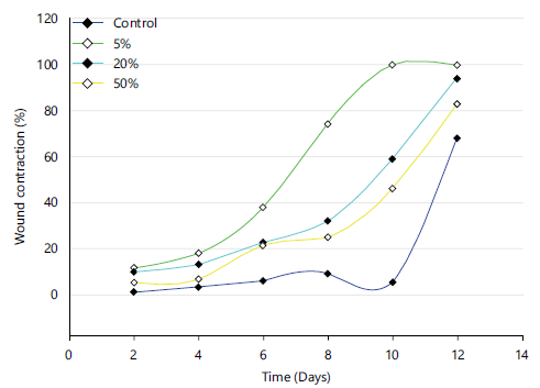
Wound contraction: Table 1 presents the percentage wound contraction observed across the different treatment concentrations over 12 days. In the control group, wound contraction was minimal during the early phase of healing, increasing only slightly from day 2 (1.1±0.9%) to day 8 (9.0±3.4%). Although a marked increase was recorded by day 12 (68±3.5%), the response was highly variable compared with the treated groups.
Animals treated with the 5% concentration showed a rapid and consistent improvement in wound closure. Wound contraction reached 11.5% by day 2 and increased progressively to 74.4% by day 8. Complete wound closure (100%) was achieved by day 10 and maintained through day 12, with no observable variation among replicates.
The 20% concentration also enhanced wound contraction compared to the control, though at a slower rate than the 5% treatment. Contraction increased steadily from 9.8±1.6% on day 2 to 32±1.7% by day 8, reaching 59±2.2% on day 10 and 94.2±1.1% by day 12.
Treatment with the 50% concentration resulted in moderate wound contraction. Initial contraction values were low during the early days (5.0±0% on day 2 and 6.6±0.9% on day 4), followed by gradual improvement to 25±0.4% by day 8. By day 12, wound contraction reached 83±8.5%, remaining lower than that observed with the 5% and 20% concentrations.
Daily rate of wound contraction (% per day): The daily rate of wound contraction, calculated at 2-day intervals, is presented in Table 2. Distinct differences in healing dynamics were observed among the treatment groups. In the control group, wound contraction progressed slowly during the early stages of healing, with low daily rates recorded between days 2 and 8 (1.10-1.50% per day). A transient decline in healing was observed between days 8 and 10 (-1.85% per day), indicating delayed or inconsistent wound repair, followed by a sharp increase in contraction rate toward the end of the study (31.35% per day).
| Table 1: | Percentage wound contraction | |||
| Contraction (%) | ||||||
| Concentration (%) | Day 2 | Day 4 | Day 6 | Day 8 | Day 10 | Day 12 |
| Control | 1.1±0.9 | 3.3±2.9 | 6.0±1.8 | 9.0±3.4 | 5.3±0.6 | 68±3.5 |
| 5 | 11.5±0 | 18±0 | 38±0 | 74.4±0 | 100±0 | 100±0 |
| 20 | 9.8±1.6 | 13.1±2.8 | 22.4±0.9 | 32±1.7 | 59±2.2 | 94.2±1.1 |
| 50 | 5±0 | 6.6±0.9 | 21.3±0.8 | 25±0.4 | 46.05±4.3 | 83±0.5 |
| Data represent Mean±SD, n = 5 and p<0.001 | ||||||
| Table 2: | Daily rate of wound contraction (% per day) | |||
| Day | Control | 5% | 20% | 50% |
| 2 | - | - | - | - |
| 4 | 1.1 | 3.25 | 1.65 | 0.8 |
| 6 | 1.35 | 10 | 4.65 | 7.35 |
| 8 | 1.5 | 18.2 | 4.8 | 1.85 |
| 10 | −1.85 | 12.8 | 13.5 | 10.53 |
| 12 | 31.35 | 0 | 17.6 | 18.48 |
| Positive values indicate active wound contraction; Zero values indicate completion or plateau of healing; Negative values indicate delayed healing or temporary wound expansion | ||||
Treatment with the 5% formulation resulted in a markedly accelerated healing response. The daily contraction rate increased substantially from 3.25% per day at day 4 to a peak of 18.20% per day between days 6 and 8. Although healing continued between days 8 and 10 (12.80% per day), the rate declined to zero by day 12, reflecting complete wound closure achieved earlier in the treatment period.
The 20% treatment group showed a steady but moderate increase in healing rate throughout the study. Daily contraction rates rose from 1.65% per day at day 4 to 13.50% per day by day 10, with a further increase to 17.60% per day between days 10 and 12, indicating sustained wound repair during the later phase of healing.
In the 50% group, wound contraction occurred at a moderate pace, with an early increase to 7.35% per day by day 6. However, healing slowed temporarily between days 6 and 8 (1.85% per day) before accelerating again during the later phase, reaching 18.48% per day by day 12.
Wound healing kinetics and overall healing performance: The progression of wound contraction over the 12 day experimental period revealed clear treatment-dependent differences in both the rate and extent of healing. Animals treated with the 5% concentration demonstrated the most rapid wound closure, achieving complete (100%) contraction by day 10, which was sustained through day 12. In contrast, the 20 and 50% treatment groups showed a more gradual healing pattern, reaching 94.2 and 83% wound contraction, respectively, by the end of the study. The control group exhibited delayed and inconsistent wound closure, with substantially lower contraction during the early and mid-healing phases.
Analysis of daily wound contraction rates further highlighted the superior performance of the 5% concentration. This group exhibited the highest healing velocity, peaking at 18.2% per day between days 6 and 8, whereas the 20% and 50% groups showed moderate but sustained increases in healing rate, particularly during the later phase of wound repair. The control group displayed irregular healing kinetics, including a transient reduction in contraction between days 8 and 10.
Area under the curve (AUC) analysis: To quantify cumulative healing efficacy, the area under the wound contraction–time curve (AUC) was calculated using the trapezoidal rule (Table 3). The 5% treatment recorded the highest AUC value (572.3 %·day), followed by the 20% (357.0 %·day) and 50% (285.9 %·day) concentrations, while the control group showed the lowest overall healing response (116.3 %·day). These findings indicate that the 5% concentration provided the most effective and sustained wound healing outcome over the study duration.
|
| Table 3: | Area under the wound contraction–time curve (AUC) | |||
| Treatment (%) | AUC (%·day) |
| Control | 116.3 |
| 5 | 572.3 |
| 20 | 357 |
| 50 | 285.9 |
Epithelialization period: The epithelialization period varied markedly among the experimental groups, reflecting differences in the rate of skin regeneration. Animals treated with the methanol extract of D. rotundifolia exhibited a reduction in epithelialization time compared to the control group, indicating enhanced re-epithelialization.
The 5% extract-treated group showed the shortest epithelialization period, consistent with its rapid wound contraction and early achievement of complete wound closure observed in the kinetic analysis. This suggests that the 5% formulation effectively promoted keratinocyte migration and proliferation, leading to faster restoration of the epidermal layer.
In contrast, the 20 and 50% extract groups demonstrated moderately reduced epithelialization periods relative to the control, although re-epithelialization occurred later than in the 5% group.
These findings indicate that higher extract concentrations supported epithelial regeneration but were less effective in accelerating the process.
The control group exhibited the longest epithelialization period, reflecting delayed epidermal regeneration and prolonged wound exposure. Overall, treatment with the methanol extract of D. rotundifolia, particularly at the 5% concentration, significantly enhanced epithelialization and contributed to improved overall wound healing outcomes shown in Fig. 1.
Results of antibacterial screening
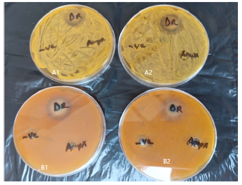
Agar Disc diffusion method: The antibacterial activity of the crude extract (DR) was evaluated using the Agar Disc diffusion method, and the results are presented in Table 4. Against the wound-sore isolate, the crude extract at a concentration of 2 mg/mL produced a clear zone of inhibition with a mean diameter of 18 mm, indicating notable antibacterial activity.
|
| Table 4: | Diameter zone of inhibition-Agar Disc diffusion method | |||
| Mean zone diameter of inhibition (mm) | |||
| Isolate | Crude extract (DR) (2mg/mL) | Ampiclox (2 mg/mL) | DMSO |
| Wound-sore isolate | 18 | Nil | Nil |
| n = 3 | |||
| Table 5: | Diameter zone of inhibition-Well-in-Agar diffusion method | |||
| Mean zone diameter of inhibition (mm) | |||
| Isolate | Crude extract (DR) (5 mg/mL) | Ampiclox (2 mg/mL) | DMSO |
| Wound-sore isolate | 20 | Nil | Nil |
| n = 3 | |||
| Table 6: | Correlation between antibacterial activity and wound healing | |||
| Group | Zone of inhibition (mm) | Day 12 contraction (%) |
| Control | 0 | 68 |
| 5% | 20 | 100 |
| Correlation coefficient (r): 1.00 and p-value: 1.00 | ||
In contrast, the reference antibiotic Ampiclox (2 mg/mL) showed no inhibitory effect against the isolate, as no zone of inhibition was observed (Fig. 2). Similarly, the negative control (DMSO) did not produce any inhibitory zone, confirming the absence of inherent antimicrobial activity from the solvent.
Well-in-Agar diffusion method: Table 5 shows the results of the antibacterial assay performed using the Well-in-Agar diffusion method. The crude extract (DR) at a higher concentration of 5 mg/mL exhibited enhanced antibacterial activity against the wound-sore isolate, with a mean zone of inhibition of 20 mm (Fig. 2). As observed in the disc diffusion assay, Ampiclox (2 mg/mL) did not inhibit the growth of the isolate, and no zone of inhibition was recorded. The DMSO control also showed no inhibitory effect.
Relationship between antibacterial activity and wound healing: Correlation analysis between antibacterial activity and wound healing outcomes showed a strong positive association between the zone of inhibition and final wound contraction. The 5% treatment, which exhibited the largest antibacterial zone (20 mm), also achieved complete wound closure, whereas the control group showed neither antibacterial activity nor comparable healing efficacy (Table 6). Although exploratory due to limited data points, this relationship suggests that the antibacterial properties of the extract may play a contributory role in enhancing wound repair.
DISCUSSION
This study provides the first scientific evidence supporting the wound-healing potential of Dissotis rotundifolia, a plant previously documented for its antiulcer activity. Soxhlet extraction of 200 g of powdered D. rotundifolia yielded 40 g of crude extract, corresponding to a 20% extraction yield. This relatively high yield suggests a substantial phytochemical content and is comparable to yields reported for other members of the Asteraceae family, reflecting effective solvent penetration and metabolite solubilization25. High extraction yields are often associated with an abundance of bioactive secondary metabolites, including flavonoids and terpenoids, which are relevant to pharmacological activity26. These findings further support the suitability of Soxhlet extraction for maximizing phytochemical recovery in pharmacognostic studies27,28.
The pharmacological rationale for investigating the wound-healing activity of D. rotundifolia is grounded in the shared pathological features of ulcers and dermal wounds, both of which involve tissue injury, inflammation, and delayed repair processes. Previous studies have demonstrated that D. rotundifolia protects gastric mucosa through mechanisms such as inhibition of gastric acid secretion, enhancement of mucin production, and antioxidant activity15,16. These mechanisms are also directly relevant to cutaneous wound repair. In vivo wound contraction results clearly demonstrated the plant’s healing potential, with the 5% extract formulation showing the greatest efficacy and achieving complete wound closure by day 10. This suggests enhanced cellular proliferation, collagen deposition, and re-epithelialization. Although the 20% formulation also promoted wound healing, the 50% concentration was comparatively less effective, resulting in only 86% wound closure.
The observed inverse dose–response pattern, in which higher extract concentrations (20% and 50%) were less effective than the 5% formulation, may be attributed to phytochemical saturation or cytotoxic effects at elevated doses. Such non-linear dose–response relationships have been reported in phytotherapeutic studies29,30 and may arise from pharmacodynamic factors including compound overload, cellular toxicity, or disruption of signaling pathways when bioactive constituents are present at supra-optimal levels. These findings highlight the importance of dose optimization in the development of effective herbal wound-healing formulations.
The wound-healing activity observed in this study is likely mediated by the same flavonoid-rich phytoconstituents responsible for the plant’s antiulcer effects16. Flavonoids are well known for their anti-inflammatory, antioxidant, and antimicrobial properties31,32, all of which play critical roles in tissue regeneration and repair. This dual pharmacological action places D. rotundifolia alongside other medicinal plants such as Aloe vera and Centella asiatica, which exhibit both gastroprotective and wound-healing properties33-36.
Furthermore, the antibacterial activity of the crude extract strengthens its therapeutic relevance in wound management. Using Agar Disc diffusion and Well-in-Agar assays, the extract produced notable zones of inhibition against bacterial strains isolated from wounds. This finding is particularly important, as microbial colonization and biofilm formation are known to delay wound healing by intensifying inflammation and promoting tissue degradation30. The antibacterial effects observed are consistent with reports on other ethnomedicinal plants that enhance wound repair by reducing microbial burden37.
The application of standardized antibacterial screening protocols recommended by the Clinical and Laboratory Standards Institute (CLSI), together with the use of clinically relevant wound isolates, enhances the translational value of these findings. Although the positive control (Ampiclox) failed to inhibit bacterial growth, possibly due to antimicrobial resistance or assay-specific conditions, the demonstrable efficacy of the D. rotundifolia extract underscores its potential as an alternative or complementary agent in wound therapy.
CONCLUSION
The findings suggest that the extract promotes wound healing partly through its antibacterial action and optimal modulation of tissue repair processes. Drosera rotundifolia therefore, represents a promising natural candidate for the development of affordable and effective wound healing formulations, warranting further investigation into its active constituents, safety profile, and mechanisms of action.
SIGNIFICANCE STATEMENT
This study provides the first scientific validation of the wound-healing potential of Dissotis rotundifolia, linking its previously reported antiulcer activity to enhanced dermal tissue repair. The findings demonstrate that a low-dose formulation (5%) optimally promotes wound contraction, re-epithelialization, and antibacterial activity, highlighting the importance of dose optimization in herbal therapeutics. By combining in vivo wound healing and antibacterial evidence using standardized methods, this research supports Dissotis rotundifolia as a promising, plant-based candidate for the development of effective and affordable wound-care agents.
ACKNOWLEDGMENTS
We wish to acknowledge the support of all the staff of Professor Dora Akunyili College of Pharmacy, Igbinedion University, Okada.
REFERENCES
- de-Luna-Gallardo, D., C. Marquez-Espriella and R. Cienfuegos-Monroy, 2024. Fundamentals of Wound Healing. In: Plastic and Reconstructive Surgery Fundamentals: A Case-Based and Comprehensive Review, de-Luna-Gallardo, D., C. Marquez-Espriella and R. Cienfuegos-Monroy (Eds.), Springer International Publishing, Switzerland, ISBN: 978-3-031-61894-9, pp: 13-22.
- Almadani, Y.H., J. Vorstenbosch, P.G. Davison and A.M. Murphy, 2021. Wound healing: A comprehensive review. Semin. Plast. Surg., 35: 141-144.
- Uberoi, A., A. McCready-Vangi and E.A. Grice, 2024. The wound microbiota: Microbial mechanisms of impaired wound healing and infection. Nat. Rev. Microbiol., 22: 507-521.
- Drabik, M. and L.H. Granicka, 2026. Advances in wound healing: Physiology, complications, the role of oxygen and innovative treatment strategies enhancing oxygenation. Biocybern. Biomed. Eng., 46: 113-129.
- Dissemond, J., J.D. Rembe, B. Assenheimer, M. Barysch-Bonderer and V. Gerber et al., 2025. Systematics, diagnosis and treatment of wound infections in chronic wounds: A position paper from WundDACH. J. Dtsch. Dermatologischen Ges., 23: 565-574.
- Graham, S.M., L. Chokotho, N. Mkandawire, M. Laubscher and S. Maqungo et al., 2025. Injury: A neglected global health challenge in low-income and middle-income countries. Lancet Global Health, 13: E613-E615.
- Sulis, G., S. Sayood and S. Gandra, 2022. Antimicrobial resistance in low- and middle-income countries: Current status and future directions. Expert Rev. Anti-Infect. Ther., 20: 147-160.
- Ahmed, L.A., Arshadul Hussain, P.A. Barbhuiya, S. Zaman and A.M. Laskar et al., 2025. Herbal medicine for the management of wounds: A systematic review of clinical studies. Infect. Disord. Drug Targets, 25.
- Albahri, G., A. Badran, A. Hijazi, A. Daou, E. Baydoun, M. Nasser and O. Merah, 2023. The therapeutic wound healing bioactivities of various medicinal plants. Life, 13.
- Yan, R., Y. Wang, W. Li and J. Sun, 2025. Promotion of chronic wound healing by plant-derived active ingredients and research progress and potential of plant polysaccharide hydrogels. Chin. Herb. Med., 17: 70-83.
- Pathak, D. and A. Mazumder, 2024. A critical overview of challenging roles of medicinal plants in improvement of wound healing technology. DARU J. Pharm. Sci., 32: 379-419.
- Shedoeva, A., D. Leavesley, Z. Upton and C. Fan, 2019. Wound healing and the use of medicinal plants. Evidence-Based Complementary Altern. Med., 2019.
- Meyer, C., H. Kreft, R. Guralnick and W. Jetz, 2015. Global priorities for an effective information basis of biodiversity distributions. Nat. Commun., 6.
- Yeboah, O.K. and N. Osafo, 2017. Review of the ethno-medical, phytochemical, pharmacological and toxicological studies on Dissotis rotundifolia (Sm.) Triana. J. Complementary Altern. Med. Res., 2: 1-11.
- Djehoue, R., A.M.O. Amoussa, A. Sanni and L. Lagnika, 2020. Phytochemical composition and antioxidant property of Dissotis rotundifolia used for malaria management in South Benin. J. Med. Plants Stud., 8: 23-29.
- >Adinortey, M.B., C. Ansah, B. Aboagye, J.K. Sarfo, O. Martey and A.K. Nyarko, 2020. Flavonoid-rich extract of Dissotis rotundifolia whole plant protects against ethanol-induced gastric mucosal damage. Biochem. Res. Int., 2020.
- Lee, J.E., J. Jayakody, J.I. Kim, J.W. Jeong and K.M. Choi et al., 2024. The influence of solvent choice on the extraction of bioactive compounds from Asteraceae: A comparative review. Foods, 13.
- Sun, W. and M.H. Shahrajabian, 2023. Therapeutic potential of phenolic compounds in medicinal plants-natural health products for human health. Molecules, 28.
- Zhang, Y., P. Cai, G. Cheng and Y. Zhang, 2022. A brief review of phenolic compounds identified from plants: Their extraction, analysis, and biological activity. Nat. Prod. Commun., 17.
- Owolabi, T.A. and P.C. Okubor, 2024. Anti-ulcer and antioxidant activities of Chrysophyllum albidum G. Don. seeds cotyledons. J. Farmasi Ilmu Kefarmasian Indonesia, 11: 260-268.
- Masson‐Meyers, D.S., T.A.M. Andrade, G.F. Caetano, F.R. Guimaraes, M.N. Leite, S.N. Leite and M.A.C. Frade, 2020. Experimental models and methods for cutaneous wound healing assessment. Int. J. Exp. Path., 101: 21-37.
- Arulprakash, K., R. Murugan, T. Ponrasu, K. Iyappan, V.S. Gayathri and L. Suguna, 2012. Efficacy of Ageratum conyzoides on tissue repair and collagen formation in rats. Clin. Exp. Dermatol., 37: 418-424.
- Umeh, V.N., E.E. Ilodigwe, D.L. Ajaghaku, E.O. Erhirhie, G.E. Moke and P.A. Akah, 2014. Wound-healing activity of the aqueous leaf extract and fractions of Ficus exasperata (Moraceae) and its safety evaluation on albino rats. J. Tradit. Complementary Med., 4: 246-252.
- Balouiri, M., M. Sadiki and S.K. Ibnsouda, 2016. Methods for in vitro evaluating antimicrobial activity: A review. J. Pharm. Anal., 6: 71-79.
- Altemimi, A., N. Lakhssassi, A. Baharlouei, D.G. Watson and D.A. Lightfoot, 2017. Phytochemicals: Extraction, isolation, and identification of bioactive compounds from plant extracts. Plants, 6.
- Owolabi, T.A. and B.A. Ayinde, 2025. Preliminary oncopharmacological evaluation of Praxelis clematidea (Griseb): Growth inhibition, cytotoxicity, and antioxidant activities. Trends Biol. Sci., 1: 295-305.
- Bitwell, C., S.S. Indra, C. Luke and M.K. Kakoma, 2023. A review of modern and conventional extraction techniques and their applications for extracting phytochemicals from plants. Sci. Afr., 19.
- Yu, X., X. Tu, L. Tao, J. Daddam, S. Li and F. Hu, 2023. Royal jelly fatty acids: Chemical composition, extraction, biological activity, and prospect. J. Funct. Foods, 111.
- Jodynis-Liebert, J. and M. Kujawska, 2020. Biphasic dose-response induced by phytochemicals: Experimental evidence. J. Clin. Med., 9.
- Lutz, W.K., R.W. Lutz, D.W. Gaylor and R.B. Conolly, 2020. Dose-Response Relationship and Extrapolation in Toxicology. Mechanistic and Statistical Considerations. In: Regulatory Toxicology, Reichl, F.X. and M. Schwenk (Eds.), Springer, Berlin Heidelberg, ISBN: 978-3-642-36206-4, pp: 1-23.
- Owolabi, T.A., E. Amodu, P. Okubor, J. Danga and D. Odiase et al., 2025. Phenylhydrazine-induced neonatal jaundice in rats ameliorated by methanol extract and the chloroform fractions of Portulaca oleracea L. (Portulacaceae). Bull. Pharm. Sci. Assiut Univ.
- Zhao, R., H. Liang, E. Clarke, C. Jackson and M. Xue, 2016. Inflammation in chronic wounds. Int. J. Mol. Sci., 17.
- Chelu, M., A.M. Musuc, M. Popa and J.C. Moreno, 2023. Aloe vera-based hydrogels for wound healing: Properties and therapeutic effects. Gels, 9.
- Matei, C.E., A.I. Visan and R. Cristescu, 2025. Aloe vera polysaccharides as therapeutic agents: Benefits versus side effects in biomedical applications. Polysaccharides, 6.
- Witkowska, K., M. Paczkowska-Walendowska, E. Garbiec and J. Cielecka-Piontek, 2024. Topical application of Centella asiatica in wound healing: Recent insights into mechanisms and clinical efficacy. Pharmaceutics, 16.
- Nitin, A., P. Kartiki, D. Pooja and D. Neha, 2025. Centella asiatica: A multifunctional phytotherapeutic approach in skin regeneration, and psoriatic inflammation control. Int. J. Pharm. Sci., 3: 2115-2128.
- Pérez-Flores, J.G., L. García-Curiel, E. Pérez-Escalante, E. Contreras-López and G.Y. Aguilar-Lira et al., 2025. Plant antimicrobial compounds and their mechanisms of action on spoilage and pathogenic bacteria: A bibliometric study and literature review. Appl. Sci., 15.
How to Cite this paper?
APA-7 Style
Owolabi,
T.A., Rafiu,
T.A., Obarisiagbon,
A.J., Dennis Olabode,
G.O., Agu,
C.C., Omoko,
A.E., Ibrahim,
O.T., Okafor,
I.C., Onimisi,
I., Abeeb,
I. (2026). Investigation of Wound Healing Potential of Methanol Exract of Dissotis rotundifolia (Sm.) Triana (Melastomatoceae). Trends in Pharmacology and Toxicology, 2(2), 60-71. https://doi.org/10.21124/tpt.2026.60.71
ACS Style
Owolabi,
T.A.; Rafiu,
T.A.; Obarisiagbon,
A.J.; Dennis Olabode,
G.O.; Agu,
C.C.; Omoko,
A.E.; Ibrahim,
O.T.; Okafor,
I.C.; Onimisi,
I.; Abeeb,
I. Investigation of Wound Healing Potential of Methanol Exract of Dissotis rotundifolia (Sm.) Triana (Melastomatoceae). Trends Pharm. Toxicol. 2026, 2, 60-71. https://doi.org/10.21124/tpt.2026.60.71
AMA Style
Owolabi
TA, Rafiu
TA, Obarisiagbon
AJ, Dennis Olabode
GO, Agu
CC, Omoko
AE, Ibrahim
OT, Okafor
IC, Onimisi
I, Abeeb
I. Investigation of Wound Healing Potential of Methanol Exract of Dissotis rotundifolia (Sm.) Triana (Melastomatoceae). Trends in Pharmacology and Toxicology. 2026; 2(2): 60-71. https://doi.org/10.21124/tpt.2026.60.71
Chicago/Turabian Style
Owolabi, Tunde, A., Toheeb A. Rafiu, Aiwaguore J. Obarisiagbon, God’s favour O. Dennis Olabode, Chinwendu C. Agu, Awesome E. Omoko, Oluwatomiwa T. Ibrahim, Izuchhukwu C. Okafor, Issa Onimisi, and Ibraheem Abeeb.
2026. "Investigation of Wound Healing Potential of Methanol Exract of Dissotis rotundifolia (Sm.) Triana (Melastomatoceae)" Trends in Pharmacology and Toxicology 2, no. 2: 60-71. https://doi.org/10.21124/tpt.2026.60.71

This work is licensed under a Creative Commons Attribution 4.0 International License.